Prcreedings of the American Chestnut Symp)sium
|
|
|
- Suzan Blair
- 5 years ago
- Views:
Transcription
1

2 Prcreedings of the American Chestnut Symp)sium Edited by William L. MacDonald, Associate Professor of Plant Pathology Franklin C. Cech, Professor of Forest Genetics John Luchok, University Editor and Clay Smith, U.S. Forest Service, NE Forest Experiment Station, Parsons, W. Va. West Virginia University Morgantown, West Virginia January 4-5, 1978
3 CONTENTS The Devastation of American Chestnut by Blight/ E. George Kuhlman l Selecting and Breeding Blight Resistant Chestnut Trees / Richard A. Jaynes Breeding of American Chestnut I Eyvind Thor Prospects for Vegetative Propagation in the Genus Castanea / Roy N. Keys The Use of Ionizing Radiation to Develop a Blight Resistant American Chestnut, CastaneaDentata, Through Induced Mutations/ Albert Dietz Chinese Chestnut Production in the Southeastern United States: Practice, Problems, and Possible Solutions I Jerry A. Payne Death of a Chestnut: The Host Pathogen Interaction / David R. M ccarroll and Eyvind Thor New Challenges in Chestnut Research/ Robert E. Phares Biological Control of Chestnut Blight in France / J. Grente and S. Berthelay-Sauret The Present Status of Chestnut Blight in Italy / Lorenzo Mittempergher American Experience with Hypovirulence in Endothia Parasitica I Sandra. Anagnostakis..., Chestnut Breeding in the United States Department of Agriculture I Frederick H. Berry The West Virginia Department of Agriculture American Chestnut Program/ J. Bruce Given and S. Clark Haynes Endothia Species as Pathogens of Chestnut and Oak / R. Jay Stipes, David N. Appel and Martha K. Roane An Evaluation of the Fungal Genus Endothia / Martha K. Roane and R. J. Stipes...., Virulence of Endothia Parasitica Isolated from Surviving American Chestnut Trees I Gary J. Griffin, John R. Elkins, Gail Tomimatsu and Frederick V. Hebard The Role of Oxalic Acid in the Pathogenesis of Endothia Parasitica I David R. McCarroll and Eyvind Thor Chestnut Callus-Cultures: Tannin Content and Colonization by EndothiaParasitica IF. V. HebardandP. B. Kaufman Effect of Some Compounds of American and Chinese Chestnut Inner Bark on the Growth of Endothia Parasitica I Safi ya Sam man, Fred M. Schell and Eyvind Thor Blight Development and Methyl-2-Benzimidazole Carbamate Levels in Bark Tissues of American Chestnut Trees Following Soil Injection ofbenomyl I John Rush Elkins, GaryJ. Griffin andr. Jay Stipes A Preliminary Report on a Method of Biological Control of the Chestnut Blight not Involving the Use of a Hypovirulent strain of Endothia Parasitica I W. H. Weidlich Insects of American Chestnut: Possible Importance and Conservation Concern / Paul A. Opler Oriental Chestnut Gall Wasp: New Nut Pest in North America/ Jerry A. Payne... : III
4 CONTENTS Research Carried Out in France Into Diseases of the Chestnut Tree / J. Grente and S. Berthelay-Sauret Some Observations on the "Hypovirulence" of Chestnut Blight in Italy/ Tullio Turchetti Pathogenicity and Sporulation of Normal and Diseased Strains of EndothiaParasitica in American Chestnut I JohnE. Elliston Testing Endothia Parasitica Strains for Vegetative Incompatibility / SandraL. Anagnostakis... IOI Frequency of Vegetative Compatibility Types ofendothiaparasitica in Two Areas of West Virginia/ William. MacDonald and Mark L. Double The Segregation of an Italian Virulent Isolate ofendothiaparasitica Into H and V Types/ N. K. VanAlfen, J. T. Bowman andj. R. Simmons Double-stranded RNA and Virus-like Particles inendothia Parasitica I J. Allan Dodds I 08 Control ofendothia Parasitica Cankers on American Chestnut Sprouts with Hypovirulent Strains IR. A. Jaynes andj. E. Elliston Pennsylvania Chestnut Blight Studies/ Barry Towers and John Longenecker Interactions of Virulent and Hypovirulent Strains of Endothia Parasitica on American Chestnuts in North Carolina/ E. G. Kuhlman Epidemiology of Hypovirulence / P. R. Day Epilogue IV
5 FOREWORD The year was 1904; the place the Bronx Zoological Park in New York City; the beginning of perhaps the greatest single natural catastrophe in the annals of forest history the discovery of chestnut blight. In less than 50 years more than 80 percent of the American chestnut trees in the eastern hardwood forests were dead; the rest were dying. A tree species that once occupied an estimated 25 percent of the eastern forest, encompassing 200 million acres of forest land, was gone. Since 1904 many futile attempts have been made to control the advance of the chestnut blight and reestablish the American chestnut in eastern forests. Frustrations have been many, rewards few. One group, the Connecticut Agricultural Experiment Station located at New Haven, Connecticut, has persisted longer than anyone in this endeavor. Recently information from Europe has stimulated renewed interest in the American chestnut problem. As one speaker at the Symposium, Eyvind Thor, so aptly stated, the history of the American chestnut has involved three stages: concern, resignation, and now, new hope for a solution. The most recent new hope is the so-called hypovirulence phenomenon that was initially observed in Italy. Hypovirulent is a term for less virulent strains of chestnut blight. In Europe, blighted chestnut trees were found with canker wounds that had healed. Canker healing was attributed to the natural occurrence of hypovirulent strains of the blight fungus that overcome or minimize the effectiveness of the more virulent strains. In this country the hypovirulent phenomenon has potential as a biological control, but there are many problems and hurdles to cross before bridging the final gap to success. During the 2-day Symposium, a total of 34 papers were presented. The first day was a general session that included historical accounts of chestnut blight and discussions of the status of selecting, breeding, and use of other techniques to produce blight resistant trees such as vegetative propagation and irradiation. The general session also included a discussion of the potential for biological control of chestnut blight in France, Italy, and the United States. A technical session on the second day provided researchers an opportunity to exchange information and ideas. The return of the American chestnut to its once significant position in the forest is doubtful. However, the sincere interest and hope remains that a solution will be found to again make it possible for American chestnut to inhabit our forest land. The broad objective of this Symposium was to exchange ideas among participants so that a more coordinated effort can be made to find a solution to the American chestnut problem. We hope this Symposium was a positive step toward achieving our long-term objective of restoring the American chestnut to the eastern hardwood forests. ACKNOWLEDGMENT We are grateful to many people for their assistance in conduct and preparation of these Proceedings. Special appreciation is extended to B. A. Breshock, J. L. Brooks, M. L. Double, D. F. Hindal, R. N. Keys, I. T. Knight, M. B. Wright, and P. S. Lotshaw. For assistance in interpretation and translation we especially thank Dr. Marilyn Bendena and Lucia Roberts. We also acknowledge the invaluable secretarial assistance of Ms Cathy L. Watson in preparation of conference materials, correspondence and manuscripts. Thanks also to the West Virginia University Office of Publications for handling the printing arrangements, and to Paul Stevenson, chief WVU Graphic Artist, and Diane Lenhart, staff artist, for their suggestions and advice on typography and design of the Proceedings.

6 Chestnut Symposium Participants (left to right). Row 1: Samuel Gingrich, Wayne Weidlich, David McCarroll, Safiya Samman, Sandra Anagnostakis, Martha Roane, Tullio Turchetti, Jean Grente, Robert Phares. Row 2: Jerry Payne, Gary Griffin, John Elkins, Richard Jaynes, Albert Dietz, Lorenzo Mittempergher, Allen Dodds, Eyvind Thor, Frankli n Cech. Row 3: Dale Hindal, Mark Double, Roy Keys, Bruce Given, Clark Haynes, John Elliston, Fred Hebard, Paul Opler, Edgar Huffman. Row 4: Clay Smith, Frederick Berry, George Kuhlman, Neal Van Allen, Peter Day, William MacDonald.
7 The Devastation of American Chestnut by Blight E. G. Kuhlman USDA Forest Service, Southeastern Forest Experiment Station, Research Triangle Park, NC ABSTRACT. A brief account of the explosive history of the classic forest tree disease, chestnut blight, in North America. The American chestnut ( Castanea dentata [ Marsh.] Borkh.) played an important role in the history of our country. Henry David Thoreau (1854) captured some of the magic of the chestnut in his book Walden. When chestnuts were ripe I laid up half a bushel for winter. It was very exciting at that season to roam the then boundless chestnut woods of Lincoln, they now sleep their long sleeps under the railroad, with a bag on my shoulder, and a stick to open burs with in my hand, for I did not always wait for the frost, amid the rustling of leaves and the loud reproofs of the red squirrels and the jays, whose half-consumed nuts I sometimes stole, for the burs which they had selected were sure to contain sound ones. Occasionally I climbed and shook the trees. They grew also behind my house, and one large tree which almost overshadowed it was, when in flower, a bouquet which scented the whole neighborhood, but the squirrels and jays got most of its fruit, the last coming in flocks early in the morning and picking the nuts out of the burs before they fell. I relinquished these trees to them and visited the more distant woods composed wholly of chestnut. These nuts, as far as they went, were a good substitute for bread. For others, nostalgia may come from recollections such as those of G. H. Hepting (1974) who said, As a boy, on cold, blustery fall and winter nights, I well remember a shivering, old Italian standing on a street corner of downtown Brooklyn before his rickety sheet metal oven-like contraption, yelling, 'Hot roasta chestnuts! Hotta roast chestnuts!' I remember the popping and crackling noises as the old fellow took off the lid to give me a nickel's worth of the sweet, hot delicious nuts.... While the fruit of the chestnut was important to man and his domesticated animals, it was even more important to the wildlife of the eastern forest. Thoreau felt smug about outsmarting an occasional squirrel or jay, but it was quite certain that wild turkeys, squirrels, jays, and other animals preferentially sought and frequently devoured this succulent fruit. The American chestnut comprised 25 percent of the eastern hardwood forest. Its natural range included over 200 million acres of land. Mature trees were feet tall with straight boles up to seven feet in diameter (Roosevelt, 1902). On good sites, open grown trees often added one inch in diameter per year and could sometimes sustain this growth for more than 50 years. Normal growth was 500 board feet per acre per year ( Holmes, 1925). Chestnut had a faster rate of growth than its associated hardwood species ( Holmes, 1925; Korstian and Stickel, 1927; Roosevelt, 1902). Chestnut wood carried man from cradle to grave, in crib and coffin. Many homes had chestnut siding, chestnut shingle roofing, and chestnut doors and paneling. Because chestnut wood was durable and rot resistant it was used for telephone poles, ship masts, railroad ties, and farm fencing. Chestnut extracts provided tannin for the leather industry. Three rather contrasting estimates of the value of chestnuts were made by the states of Pennsylvania, North Carolina, and West Virginia from 1909 information. Detwiler (1912) estimated 7.6 million acres of forest land in Pennsylvania with 21 percent in chestnut timber. He allowed two poles, four railroad ties, and two cords of wood per acre at a value of two dollars per pole, 33 cents per tie, and one dollar per cord. Total timber value was $55 million. Nut crop, orchard, park, and shade trees had an estimated worth of another $15 million. Buttrick (1925) estimated 31/2 billion feet of standing chestnut in North Carolina in an area of approximately 7.6 million acres. Only 50 percent of this timber was accessible to lumbermen. For the accessible portion the estimated value was 11/2 dollars per thousand bd. ft. for saw timber and 121/2 cents per cord for cordwood. Total value of the chestnut was therefore only $2.5 million in North Carolina. In West Virginia there was an estimated ten billion bd. ft. of standing chestnut worth 21/2 dollars per thousand bd. ft. for a total value of $25 million ( Giddings, 1912). West Virginia reported that one railroad station shipped 155,000 lbs. of chestnuts in 1911 ( Giddings, 1912). The U.S. Forest Service's estimate for chestnut timber cut in 1909 was $20 million ( Detwiler, 1912). H. W. Merkel (1905), chief forester and constructor of the New York Zoological Society, discovered the chestnut blight disease in the Bronx parks. "During 1904," he reported, "an epidemic of a fungus disease has occurred throughout the parks of this Borough, which but for the fact that it was confined to a single species of tree, might have overshadowed in deadliness and rapid spread all other enemies of tree life." Merkel ( 1905) obtained an emergency appropriation of $2,000 to treat the affected trees. With that money he trimmed out the
8 disease from 438 individuals. In spite of this effort he reported that 98 percent of all chestnuts in Bronx parks were infected in The fungus was highly virulent; in one case only 21 days elapsed between the first symptom and the girdling of a 4-in. stem. Merkel (1905) reported the physical condition of the tree had no effect on the fungus. The disease was equally as frequent on young nursery specimens, sprouts, and young trees feet tall as it was on old patriarchs feet in circumference. It should be noted that although the disease frequency was similar on all trees, the response of individual trees and even limbs on the same tree varied greatly. Generally, infections rapidly girdled smooth-barked branches. However, girdling of rough-barked limbs took from one to ten years with an average of three to four years ( Gravatt, 1925). A botanist, W. A. Murrill (1906), of the New York Botanical Gardens, reported that inoculation studies indicated infection probably took place only through wounds, which unfortunately were all too common on chestnut. The causal fungus was briefly called Diaporthe parasitica but was soon named Endothia parasitica ( Murr.) P. J. & H. W. And. (Shear, et al., 1917). Undoubtedly the blight had entered this country before 1904 when Merkel found the affected trees in the Bronx, but it was a few years before the source of the fungus was determined. Initially the sudden outbreak was attributed to severe drought conditions that made chestnut susceptible to an otherwise innocuous fungus. Evidence was soon presented that the disease was introduced from a foreign country. Observations in the eastern United States had indicated Chinese and Japanese chestnuts had more natural resistance to the disease than did the American chestnut ( Shear et al., 1917). If the host and pathogen evolved together there would have been selection pressure on both and some resistance would have occurred. Thus natural resistance in Asiatic chestnuts indicated an Asian origin for the pathogen was likely. In the fall of 1912 diseased chestnut material from Agassiz, British Columbia, proved to contain E. parasitica ( Shear et al., 1917). Chestnut was not native to British Colubbia and the Agassiz planting contained stock of American, European, and Asian origin. Although all the trees were ordered from American nursery firms, the planting supervisor remembered the Asian species were shipped to Agassiz in the original wrappings which consisted of distinctive Asian mats and casings. In 1913, Frank N. Meyer, an agricultural explorer, found the fungus in China on native chestnuts and subsequently he also found it in Japan. Isolates from these specimens caused symptoms identical to those caused by isolates from the United States on American chestnuts ( Shear et al., 1917). Most conditions appeared to favor the pathogen. A highly susceptible host evenly distributed through its range, a favorable climate, no natural barriers to limit spread, and two abundant spore forms provided an efficient means of spread. The disease spread rapidly from the New York City area. In 1909, the USDA indicated that most chestnuts within 30 miles of New York City were infected and scattered disease centers were present in Pennsylvania, Virginia, Maryland, Connecticut, and Rhode Island (Metcalf and Collins, 1909). Two years later, the main disease center was 150 miles south and miles north and west of New York City, with scattered centers up to 120 miles in advance of this main center (Metcalf and Collins, 1911). The rate of spread of the main disease center was given as ten miles per year. However, spot infection centers developed up to 150 miles from the leading edge. One such large center involving three counties in North and South Carolina was first noticed in 1923, but its size suggested it started at least as early as 1912 ( Gravatt, 1925). These spot infections were thought to result from spores carried on birds; however, movement of chestnut products was not restricted and probably contributed to the rapid dissemination (Detwiler, 1914; Gravatt, 1935). By 1950 the blight occurred on more than 80 percent of the trees throughout the range of American chestnut. Efforts to control chestnut blight were started by Merkel, the discoverer of the disease. He attempted control by cutting out the affected tissue and by spraying with Bordeaux mixture (Merkel, 1905). Neither method provided control in New York City. Regional efforts to eradicate the disease were made from Metcalf and Collins (1911) of the USDA, Bureau of Plant Industry located 14 spot infection centers within a 35-mile radius of Washington, D.C., in All infected trees in these centers were felled, and the bark and brush were burned on the stumps. In 1911 no new cases of the disease were reported in the 35-mile zone (Metcalf and Collins, 1911). F. C. Stewart (1912) of the New York Agricultural Experiment Station analyzed Metcalf and Collins' work in a paper he presented at the Pennsylvania Chestnut Tree Blight Conference in His first criticism was "... there was no check treatment and experimenters are agreed that experiments without checks have little value." Secondly, Stewart visited two centers of infection within Metcalf and Collins' "immune zone." One tree over 3 ft. in diameter was in advanced decline and must have been infected for several years, including the time Metcalf was stating that the area was apparently free from the disease. Finally, Stewart visited two treated areas and in one found the fungus in bark that had not been removed from the stump. A nearby tree also was infected. Stewart correctly predicted that eradication would not be effective in controlling the disease. Although his speech was low key and scholarly it became the target of other speeches at the Pennsylvania Conference. The prevailing mood was patriotism and was exemplified by Pennsylvania's willingness to invest $275,000 in an effort to stop the disease. That mood and effort were not
9 slowed by Stewart. Although others echoed his sentiments they veiled their criticisms, whereas Stewart's title "Can Chestnut Bark Disease be Controlled?" was immediately answered negatively in his text. When the Pennsylvania effort was abandoned two years later, it was suggested the disease had been slowed by five years through the effort (Sargent, 1914). The failure to eliminate the disease by eradication in New York, Pennsylvania, and the District of Columbia can be understood on the basis of the large numbers of trees involved. Furthermore, evidence from several outbreaks in ornamental and orchard plantings in the western United States proved that even limited infestations were impossible to eradicate. At the Agassiz, B.C., site, all infected trees were destroyed in 1912; however, the disease appeared on other trees in 1934 ( Gravatt, 1935). In Gunter, Oregon, the disease was found on two trees in 1929; these trees were cut and burned. However, in 1934 the fungus was still active on one stump a foot below ground ( Gravatt, 1935). In California, the disease persisted from 1934 until at least 1945 in spite of meticulous eradication and sanitation efforts in the orchards on an annual basis ( Milbrath, 1945). The second major emphasis in control has been on efforts to breed chestnuts that are resistant to blight. Because four other papers are on the subject of resistance, it is sufficient to indicate that the earliest papers (Murrill, 1906) broached the subject of resistance and that it has remained a promising hope. The American chestnut has been devastated by blight. This once prominent species has reverted to a very minor role in the eastern forests. The hopeful leads for the revitalization of this species will be the topic of other papers. LITERATURE CITED Buttrick, P. L CHESTNUT IN NORTH CAROLINA. In Chestnut and the Chestnut Blight in North Carolina. N.C. Geol. and Econ. Surv. Econ. Pap. 56. p Detwiler, S. B THE PENNSYLVANIA PROGRAMME. Pa. Chestnut Blight Conf., Harrisburg. p Detwiler, S. B OBSERVATIONS OF SANITATION CUTTING IN CONTROLLING THE CHESTNUT BLIGHT IN PENNSYL- VANIA. Final Rep. Pa. Chestnut Tree Blight Comm., 1913, Harrisburg. p Giddings, N. J (Untitled report on chestnut blight in West Virginia.) Pa. Chestnut Blight Conf., Harrisburg. p. 26, Gravatt, G. F THE CHESTNUT BLIGHT IN NORTH CAROLINA. In Chestnut and the Chestnut Blight in North Carolina. N.C. Geol. and Econ. Surv. Econ. Pap. 56. p Gravatt, G. F CHESTNUT BLIGHT IN CALIFORNIA. I. DEVELOP- MENT OF THE DISEASE. Mon. Bull. Calif. Dept. Agr. 24: Hepting, G. H DEATH OF THE AMERICAN CHESTNUT. J. For. Hist. 18: Holmes, J. S FOREWARD. In Chestnut and the Chestnut Blight in North Carolina. N.C. Geol. and Econ. Surv. Econ. Pap. 56. p Korstian, C. F. and P. W. Stickel THE NATURAL REPLACEMENT OF BLIGHT-KILLED CHESTNUT. USDA Misc. Circ p. Merkel, H. W A DEADLY FUNGUS ON THE AMERICAN CHEST- NUT. N.Y. Zool. Soc. 10th Ann. Rep. p Metcalf, H. and J. F. Collins THE PRESENT STATUS OF THE CHESTNUT BARK DISEASE. USDA Bull p Metcalf, H. and J. F. Collins THE CONTROL OF THE CHESTNUT BARK DIS- EASE. USDA Farmers' Bull p. Milbrath, D. G BUREAU OF PLANT PATHOLOGY. Calif. Dept. Agr. Rep. 34: Murrill, W. A FURTHER REMARKS ON A SERIOUS CHESTNUT DISEASE. J. N.Y. Bot. Gard. 7: Roosevelt, T. R MESSAGE FROM THE PRESIDENT OF THE UNITED STATES, TRANSMITTING A REPORT OF THE SECRE- TARY OF AGRICULTURE IN RELATION TO THE FOR- ESTS, RIVERS, AND MOUNTAINS OF THE SOUTHERN APPALACHIAN REGION. Govt. Print. Off., Washington, D.C. 210 p. Sargent, W LETTER OF TRANSMITTAL. THE COMMISSION FOR THE INVESTIGATION AND CONTROL OF THE CHESTNUT TREE BLIGHT IN PENNSYLVANIA. Final Rep. Pa. Chestnut Tree Blight Comm., 1913, Harrisburg. p Shear, C. L., N. E. Stevens, and R. J. Tiller ENDOTHIA PARASITICA AND RELATED SPECIES. USDA Bull p. Stewart, F. C CAN THE CHESTNUT BARK DISEASE BE CON- TROLLED? Pa. Chestnut Blight Conf., Harrisburg. p Thoreau, H. D WALDEN. 357 p. Houghton, Mifflin & Co., Boston.

10 Selecting and Breeding Blight Resistant Chestnut Trees Richard A. Jaynes Connecticut Agricultural Experiment Station, New Haven, CT ABSTRACT. Background information of the species of Castanea, their identification, flower morphology, and cross-pollination techniques are important to understanding chestnut breeding. The future of chestnut breeding depends on several factors, but in the long run breeding will play a role in the continued existence of this species. "Work of this kind (breeding) is extremely valuable and, although slow in yielding results, may eventually prove to be the only means of continuing the existence in our land of a greatly esteemed tree." I DENTIFICATION There is often confusion over identification of the various chestnut species. Any one trait is often inadequate to distinguish the species, but taken together, leaf shape, leaf margin, hairs on the leaf undersurface, twig color, and bud shape are useful key characteristics ( Fig. 1). Whenever we have reports of large American chestnuts, 10-in. diameter breast height ( dbh) or larger, we attempt to have a twig and leaf sample sent to us to confirm identification. Usually such reported trees are not the native chestnut. Confirmation by mail forestalls many unrewarding field inspection trips. These words were written by Arthur H. Graves in 1914, who worked with chestnut from 1911 to 1962 and began breeding chestnut trees in Little did he realize how slow progress might be; however, it was and still is a valid approach that has proved successful with agronomic, horticultural, and forest crops. Some background on the species of Castanea, their identification, flower morphology, and the techniques of cross-pollination are important to understanding the breeding that has been and can be done. Much of what I have to say is a review of information already in the literature ( Dierauf, 1977; Jaynes, 1969, 1972; Keys et al., 1975). SPECIES There are 13 species of chestnut native to the north temperate zones of Asia, Europe, and the United States. Only the American chestnut (Castanea dentata [ Marsh.] Borkh. ) was a notable forest tree. Several of the species are small trees or shrubs of minor economic importance. The European ( Gastanea sativa Mill.), Chinese (C. mollissima Bl.), and Japanese (C. crenata Sieb. & Zucc. ) chestnuts are all valued primarily as nut producers and only secondarily for wood. Although the latter three species can grow to large size they seldom develop the straight clear bole that was characteristic of forestgrown American chestnut. Two species, the Chinese and Japanese chestnut, have high levels of resistance to the chestnut blight fungus, Endothia parasitica ( Murr. ) P. J. & H. W. And. The resistant species and the parasite evolved together, whereas the American and European chestnut species evolved without the selective pressure of the chestnut blight fungus and, therefore, were genetically unprepared when exposed to the disease organism. Figure 1. Twigs and leaves of three species of chestnut. Left, the Chinese chestnut twigs have a li ght, yellowish-buff winter twig color; there are simple hairs at the tip of the twig and the leaf is broad. Center, the Japanese chestnut has rounded buds and a leaf that is narrow and bristle tipped with a crenate margin. Right, the American chestnut leaf has an angular base compared to the two Oriental chestnuts and the leaf margin is more dentate. The size of leaf varies and is not critical in identification. FLOWERS Chestnuts are monoecious, that is, male and female flowers are separate but both occur on the same tree. They are borne on the current year's growth. Two types of inflorescence are found: the unisexual male catkins, located on the lower parts of the shoot, and the bisexual catkins toward the
 normally contains three chestnuts, whereas the chinkapins are characterized by one nut.")
11 terminal end of the shoots ( Fig. 2 ). Pistillate or female inflorescences appear singly or in clusters of two or three at the base of the bisexual catkins. The bur or involucre of the true chestnuts ( American, Chinese, European, and Japanese) normally contains three chestnuts, whereas the chinkapins are characterized by one nut. Flowering is late compared to most temperate tree species and occurs after the first leaves have fully expanded. There is variation according to species, clone, and season. The male catkins shed pollen first, then in a few days the styles of the pistillate flowers spread, and last the male flowers of the bisexual catkins open. Chestnuts rarely selfpollinate. They are predominantly a wind-pollinated species with insects playing a minor role in crosspollination. Figure 2. Chestnut flowers. The male or staminate catkins have started to shed pollen. The developing burs are at the base of the more distal, bisexual catkins. The styles are beginning to spread. Pollen will not be shed from the male flowers on the bisexual catkins for several days. CONTROLLED CROSSES Female flowers are not receptive until five days after pollen shedding by the male catkins begins (anthesis ); best results with controlled crosses are obtained when pollinations are made days later. There is a temptation, because of the spread styles on the pistillate flowers, to not wait long enough before pollinating. Female flowers to be pollinated are generally isolated in water-proof paper bags just prior to their period of receptivity. Crosses are made with fresh catkins that have been bagged and are shedding pollen or with stored pollen. Pollen can be dried and stored frozen for a year. Controlled crosses are not especially difficult but are time consuming. One alternative, which has value in some situations, is to take advantage of the self-sterility of chestnut trees. Two different isolated chestnut clones will normally intercross. Theoretically, large numbers of hybrid seed could be obtained from isolated two-clone plantings. BREEDING CHESTNUT A true awareness of the value of the American chestnut as a nut tree began to develop in the late 1800's. Some efforts were made at selecting outstanding native clones, and European selections, namely the 'Paragon,' were grafted on to native chestnut sprouts. Luther Burbank in California and Walter Van Fleet in Maryland had begun to hybridize the different species to produce better nutbearing selections. A significant domestic chestnutorchard industry was in the making when the chestnut blight fungus struck. The American and European trees were killed but Japanese chestnuts and some of their hybrids were resistant. The breeding for a forest tree with characteristics of the American chestnut plus the trait of resistance to E. parasitica was begun in earnest by Flippo Gravatt and Russell B. Clapper of the USDA in Arthur Graves started his chestnut breeding in 1929 and these efforts have been continued by. Nienstedt, myself, and others at the Connecticut Agricultural Experiment Station. Numerous other individuals and institutions also have played a role in the selection and breeding of hybrid chestnuts. Hybridization among the species is not difficult. Indeed, the numerous F 1 crosses made among ten species illustrate relative free compatibility, indicating the potential for gene exchange among the species. Breeding for a blight resistant forest tree has concentrated on the use of three species: the American, Chinese, and Japanese chestnut. Early workers hoped that F 1 hybrids would meet their needs but as these trees matured it became apparent that, despite good form and vigor, they lacked adequate field resistance to the blight. Numerous second and third generation crosses of various combinations of the three species were tested but no one cross has yielded the desired result. In fact, there are few if any single, hybrid trees of large size that can be pointed to as growing like an American chestnut and being blight resistant. One of the most promising and highly publicized hybrids was the `Clapper' chestnut which succumbed in 1976 after carrying a latent infection for many years. Failure to achieve the desired result does not condemn the methods used. Three major handicaps
12 to the breeding work are: 1) the lack of a satisfactory means to screen young seedlings for blight resistance, 2) no ready means to vegetatively propagate and thus test selections on their own roots, and 3) populations of hybrids have been too small to obtain the desired segregation. There appears to be linkage of traits for poor form with blight resistance and, conversely, good form and vigor with blight susceptibility. Breeders have failed to recognize the need for, or have been unable to grow, large populations of hybrids. The largest hybrid chestnut planting is on the Lesesne State Forest in Virginia. Through the financial aid of Mrs. Arthur Valk and the cooperation of the Virginia Division of Forestry and the Connecticut Agricultural Experiment Station over 10,000 hybrid chestnut seedlings were planted between 1969 and Survival has been good. The oldest plantings have formed a closed canopy, with trees up to 30 feet tall and 4 in. dbh. A few of the trees in this planting are from controlled crosses, but most of them are from single parent selections such as the 'Clapper' chestnut. In many cases the seed parent was in a planting of selected hybrids and so the offspring are the products of natural crossing of selected hybrid trees. They represent third to fifth generation selections. At this early date only 4 percent of the seedlings at the Lesesne State Forest promise to be good timber types. Little blight infection has occurred among these hybrids as contrasted to a nearby planting of American chestnut seedlings. These hybrids represent the best gene source in the country for future selection and breeding of a blight resistant forest tree. With substantial effort, it should still be possible to develop clonal selections and even relatively true breeding lines of blight resistant, timber chestnuts for the eastern United States. THE FUTURE Whether the selection and breeding of hybrid chestnut trees should continue with vigor depends on many factors, not the least of which is the practical control of the chestnut blight fungus. Obviously, if American chestnut can again be grown, species-hybrid chestnut trees are not needed for the forest. However, even if we should be able to grow the native chestnut, we need to recognize that, as much as the tree has been idolized, it is not perfect and selective breeding within the species may well be warranted. Certainly there is considerable variation within the species for numerous characteristics. It also is possible that the hypovirulent strains could exert effective control on trees which we now consider to have inadequate field resistance, such as the 'Clapper' chestnut. Should this be true then selected hybrids at Lesesne might have immediate value. Finally, regardless of the outcome of biological control of the blight, there is interest and demand for ornamental and nut-bearing chestnuts for the home and orchard. Chinese chestnut seedlings supply some of this need, but clonally propagated selections would be more satisfactory. Hybrids, present and future, would play an important role, such as third and fourth generation selections of the Chinese-Seguin (C. mollissima-c. seguinii) hybrids. These offer the advantages of the blight resistant Chinese chestnut with prolific, precocious bearing on trees of small stature. Chestnut breeding is in a state of flux. The future will depend on many factors: the spread of the newly introduced gall wasp (Payne, 1979), improvement of vegetative propagation techniques, rapid screening of seedlings for disease resistance, and, most important, on the outcome of present biological control efforts. Breeding, in the long run, will play a role in the continued existence of this greatly esteemed tree. LITERATURE CITED Dierauf, T. A CHESTNUT RESEARCH IN VIRGINIA. Ann. Rept. Northern Nut Growers Assoc. 68: (in press(. Jaynes, R. A GENETICS OF CHESTNUTS. USDA Forest Serv. Res. Pap. WO-17, 13 pp. (revision in press(. Jaynes, R. A BREEDING IMPROVED NUT TREES. In: Handbook of N. American Nut Trees. Ed. Jaynes, Northern Nut Growers Assoc., Knoxville, TN. Keys, R. N., F. C. Cech, and W. MacDonald PERFORMANCE OF CHINESE AND HYBRID CHESTNUT AFTER TWENTY YEARS. Ann. Rept. Northern Nut Growers Assoc. 66: Payne, J. A ORIENTAL CHESTNUT GALL WASP: NEW NUT PEST IN NORTH AMERICA. American Chestnut Symposium Proceedings, West Virginia University, Morgantown, WV.
13 Breeding of American Chestnut Eyvind Thor Department of Forestry, Wildlife and Fisheries, The University of Tennessee, Knoxville, TN ABSTRACT. The low level of variation in blight resistance among native American chestnut trees has discouraged tree breeders from working with intraspecific hybridization in this species. If a less virulent fungus becomes established, chances are much better that the variation present will be sufficient to develop trees with adequate resistance. A recurrent selection breeding program is outlined and suggestions are made for cooperation in future breeding efforts. The history of chestnut blight in America and the efforts to control the blight have been discussed in some detail by several speakers in the General Session. It may be concluded that we have been through two stages in history of the disease. The first stage was a "period of great concern" starting with the outbreak of the disease and ending with World War II. After WWII, interest in the American Chestnut (Castanea dentata [ Marsh.) Borkh.) declined rapidly and by 1960, when the U.S. Forest Service dropped its research project, we reached the bottom of the second stage, which may be called "the period of resignation." This period lasted until about 1970 when conditions appeared more favorable and new research was started. The papers presented at this Conference give the background for this renewed interest in American chestnut, and the large number of participants give hope that we are now in the third and final stage. May this stage go down in history as "the period of solutions." Biological control of the blight using hypovirulent strains is the main cause for renewed interest in the American chestnut. If further research with hypovirulence results in development of practical control methods in the field, there may be new opportunities for tree breeders interested in this species. Rather than having to breed for just a single characteristic, resistance to blight, the chestnut breeder may also consider characteristics such as nut and timber production. However, until such methods have been developed it is still prudent to concentrate on the single objective of increasing the amount of resistance in the American chestnut. VARIATION IN BLIGHT RESISTANCE The apparent lack of resistance to the blight fungus (Endothia parasitica [ Mum] P. J. & H. W. And.) in the American chestnut made breeders look to other Castanea species for genes controlling resistance. A classic plant breeding program for disease resistance using interspecific hybridization and back crossing resulted in some trees with a high degree of resistance but poor growth form while other trees had good form but only intermediate resistance. Apparently, it is very difficult to combine high resistance and good growth form. The lack of unqualified success in the hybrid breeding program made some tree breeders, professionals as well as amateurs, take a second look at the remaining population of American chestnuts. Although the picture in general was very discouraging there were numerous reports of large surviving native trees. These trees were not escapees; old cankers gave evidence that they had survived repeated attacks. The limited amount of resistance in American chestnut may, at least theoretically, be enhanced by use of ionizing radiation. Some new varieties of agronomic species have resulted from radiation breeding, but the conditions conducive to success with small grains are not found in American chestnut. Seed of the American chestnut are limited in supply and very costly. Also, individual seedlings must be transplanted to large field plots where they must be maintained for several years before they start flowering. Since most mutants are recessive, large second-generation populations must be grown and tested to have a reasonable chance of finding a resistant mutant. Such large chestnut plantations would be extremely expensive to establish and maintain. While ionizing radiation breeding may be successful, work should be continued with the selection-hybridization program. Considering the limited resistance in the American chestnut, selection would have to be carried out for a number of generations to obtain trees with a substantial amount of resistance to the present virulent strains. If, however, hypovirulent fungal strains can be established which will eliminate the virulent strains, it may be possible to breed trees with sufficient resistance for protection against these new strains. The new and hopefully much lower level of resistance needed in the future may not be known for several years. However, this time period will be relatively short compared to the time requirements of a selection breeding program. The present lack of a standard for the amount of resistance needed should not discourage an aggressive breeding program; it is indeed difficult to comprehend how American chestnut trees with an excess amount of resistance to the blight can be produced!

14 SELECTION OF SUPERIOR PHENOTYPES The first step in a program of recurrent selection is the selection of the base population. Superior phenotypes of American chestnut are not common; large surviving trees are indeed so unusual that they are identified and talked about by landowners, foresters and hikers. Newspaper publicity regarding the breeding program will usually bring a number of letters with information on surviving trees. Most of them will lead to someone's backyard and an Oriental chestnut tree. However, occasionally American chestnut trees are found in forests and fields. The breeder may not want to include any and all surviving American chestnut trees in his base population. He will want to select those showing promise of the largest amount of resistance. For that reason only relatively large trees, at least 10 to 12 inches in diameter, should be considered. Such trees have had ample opportunity to become infested through cracks and wounds in the bark. Also, they have been screened for resistance both as juvenile and mature trees. Evidence of old cankers on the stem does not disqualify a tree for use in a breeding program; on the contrary, a tree that has succeeded in stopping fungal growth and started healing over the wounds may have greater breeding value than one which has been successful in avoiding infection. Further evaluation of a candidate tree, especially one with no signs of infection, may be obtained by artificial inoculation. This test may be made directly on the tree or on excised branches in the laboratory. Papers in the Technical Session discuss some of these methods. within a couple of years and tend to have heavy annual nut crops ( Fig. 2 ). CLONAL TEST AND BREEDING ORCHARD In the University of Tennessee breeding program, the last step in selection of the base population and the first step toward production of progenies is combined in a clonal breeding orchard. Ramets of the selected trees are established by grafting physiologically mature scion wood to potted Chinese chestnut seedlings. This operation is carried out in the greenhouse during the winter with dormant scion wood and actively growing understock. After one or two years in the shadehouse the grafted trees may be transplanted to the breeding orchard. This orchard will then serve two purposes; natural infection will screen out the less resistant clones and the remaining clones will be used for breeding purposes. More efficient clonal tests can be established by using rooted cuttings. However, only juvenile cuttings will root easily; to date we have only obtained callus formation on mature wood ( Fig. 1). The disadvantage of using juvenile shoots is that several years are needed before the surviving ramets start producing flowers. Ramets produced by grafting of mature shoots will start flower production Figure 2. Scion wood taken from American chestnut trees selected for apparent resistance to the blight are grafted on Chinese understock. This ten-year-old ramet has an abundant crop of chestnuts.

15 PROGENY TESTING Two types of progenies are produced in the breeding orchards. They are the result of either open or controlled pollination (Fig. 3). Open pollination results in half-sib families while full-sib families can be obtained from controlled pollination. There are some important differences between them. Half-sib families are comparatively inexpensive, but half-sib progeny tests will only give information on general combining ability. If the progenies of a given parent on the average have a high degree of resistance, this tree has good general combining ability for this trait. Due to the tedious task of control pollination in chestnut species the full-sib families are expensive to produce, but progeny tests with full-sib families will, in addition to general combining ability, give information on specific combining ability. If two parents produce progenies with more resistance than that expected based on their general combining ability they have good specific combining ability. Also, with controlled pollination, maternal effects can be assessed. To obtain valid data of specific or general combining abilities it is necessary to use a suitable experimental design in the establishment of the progeny test. Several such designs are being used for other forest tree species and may be adapted to use in progeny tests of American chestnut. However, due to the variable number of progenies available and the expected high mortality the simpler designs offer greater flexibility. RECURRENT SELECTION The main purpose of the progeny test may be to evaluate the parents in the breeding orchard. Parents with poor combining ability can then be removed from the orchard. However, the progeny test also provides material for second generation selection. Progenies of poor families will be avoided, while the best phenotypes within the best families are selected for further breeding. Timing of this selection can be very important. Since the amount of resistance should increase with each generation of selection it is desirable to maintain a very short rotation age. Theoretically this rotation age should be equal to the time required for the trees to start flowering. In American chestnut this may be as little as five to six years. However, it is of little use to turn over generations if a meaningful selection differential is not maintained. By prolonging the rotations to about ten years natural selection will have reduced the task of further evaluation with artificial inoculation and the selected trees will be of sufficient size and age to provide all the flowers needed for production of the next generation. Another problem encountered in a program of recurrent selection is that of inbreeding. This problem may appear very early in the breeding program if the number of parents used in the first generation is small ( less than surviving clones in the breeding orchard) and if second generation selection is carried out in open-pollinated progeny test plantations. Both mass selection and selection of individuals within open-pollinated families may result in a severe reduction of variation because most of the trees selected may have one parent in common, a tree with exceptionally good general combining ability. The use of full-sib family test plantations and selection of progenies with different pedigrees may be needed to maintain the variation necessary for recurrent selections. CONCLUSIONS A recurrent selection breeding program as outlined above may appear to be relatively simple. There are, however, several practical problems which have one thing in common they require large investments of time and money. Considering the large commitment of personnel and funds for a long period of time it is easy to understand why most forestry research organizations do not have breeding programs with American chestnut. At the University of Tennessee we have been engaged in this type of research for about 15 years, but limited funding has severely restricted the quantity of suitable breeding material. It is curious that today an obscure fish or plant may stop projects worth hundreds of millions of dollars. The only requirement is that it be put on a list of endangered species. The American chestnut is, of course, too ubiquitous for such consideration;
16 it must be almost as common as the passenger pigeon was once. To save the passenger pigeon would have required a regional effort and, likewise, the chestnut blight is not a problem of any specific state and should not be the responsibility of any one state to solve. The resources needed to solve the problem require input from the federal government and cooperation among all participating institutions. In this respect, the renewed interest by the U.S. Forest Service in the American chestnut is very gratifying. A longterm commitment by the largest forestry research organization in the world should provide the leadership, coordination, and continuity needed to carry out a meaningful program. For many years breeding efforts with American chestnut were carried out by enthusiastic amateurs at their own expense and often in their own backyards. Even though their efforts often were based more on faith than science and the reaction of professional breeders often was condescending they succeeded in establishing some plant material which should be of use in the future. If additional professional breeding programs with the American chestnut get started, it is essential that the amateur breeders be included; they can provide valuable services, particularly in selection and testing. Prospects for Vegetative Propagation in the Genus Castanea Roy N. Keys Division of Forestry, West Virginia University, Morgantown, WV ABSTRACT. Literature concerning the vegetative propagation of chestnut by grafting, rooting cuttings, layering, and tissue and organ culture is reviewed. It is concluded that attempts to develop a system of propagating desirable chestnut clones should be continued. The most promising techniques are concluded to be nut grafting, rooting cuttings and tissue and organ culture. In 1925, the United States Department of Agriculture at Beltsville, Maryland, instituted a program to breed chestnut trees which would be resistant to the blight fungus (Endothia parasitica [ Murr.] P. J. and H. W. And.) ( Saucier, 1973). Four years later the Brooklyn Botanical Garden started a similar program, which was later continued at the Connecticut Agricultural Experiment Station at Hamden, Connecticut (Jaynes, 1972). Meanwhile, a search for resistant American chestnut ( Castanea dentata [ Marsh] Borkh), was being conducted by various state and federal agencies. While neither program has been completely successful, there are a small number of residual trees which may have genes for partial resistance, and there are several hybrids which show some resistance and have good form. Success in either of these programs will necessitate the development of a practical technique for vegetative propagation. The fortunate gene combination will rarely occur and the genus Castanea is generally self-sterile (Clapper, 1954), so the establishment of pure breeding lines would be difficult or impossible. Therefore vegetative propagation is the only alternative for the multiplication of desirable trees. Vegetative propagation can be accomplished by grafting, rooting cuttings, layering, or the relatively new procedure of growing plantlets through tissue culture. Grafting Grafting has generally been more successful as a means of propagating chestnut than layering or rooting cuttings. Grafting consists of placing a twig ( scion) from the resistant plant into another seedling or tree ( stock plant). The two grow together after a period of time, creating a new plant. Splice, whip, cleft, and side grafts have been used successfully in bench grafting, or grafting in the greenhouse (Fig. 1). The splice graft is simplest and seems to be the most effective ( Nienstadt and Graves, 1955). This type of graft can also be used in field grafting such as is done on stock plants in the seedbed or the seed orchard (Jaynes, 1972). Mature trees can be topworked using the veneer crown graft ( Nienstadt and Graves, 1955) (Fig. 2) or bark graft ( McKay and Jaynes, 1969). The best time for scion collection is February or March (Nienstadt and Graves, 1955). The scion should be cut to 12 inches in length and stored in nearly dry peat moss at F in sealed plastic bags (Jaynes, 1969). Field grafting should be done when the leaves of the rootstock are mature to avoid frost damage (Nienstadt and Graves, 1955). Nienstadt and Graves (1955) recommend grafting onto well-established stock plants. Park (1967), however, grafted onto juvenile tissue of Japanese chestnut (C. crenata Sieb. & Zucc.) with some success. The rootstock consisted of newly germinated seeds. The graft was made onto the epicotyl ( stem) which had at least four mature leaves. The

17 scions were either newly elongated shoots of mature trees or epicotyls with four mature leaves. A major problem associated with the grafting of chestnuts is incompatibility between the rootstock and scion. This is caused when the scion does not make a completely successful union, and can be compared to the rejection of a transplanted organ in humans. This problem can be alleviated by choosing the proper rootstock. McKay and Jaynes (1969) recommend using seed collected from the variety being grafted as a source of stock plants for Chinese chestnut (C. mollissima Bl. ). The degree of success in grafting hybrid scions was affected by the species of rootstock, with no apparent pattern to compatibility ( Stairs, 1964 ). Morris reported that chinquapin would accept almost any species of chestnut, but appears to have a dwarfing affect on the scion ( Kains and McQuesten, 1967). Incompatibility is probably genetically controlled, as indicated in Park's (1967) study. In three trials using Japanese chestnut rootstock and scions of three clones, success was 0, 10, and 70 percent. Another problem with grafting is its prohibitively high cost. This factor alone eliminates grafting as a means of mass-producing desirable clones. Only in a situation where the final product yields a rapid return and high price (ie., commercial nut production), or the goal is preservation of desirable germ plasm (ie., seed orchards), would grafting be economically feasible. One technique which might alleviate these two problems is nut grafting. Moore (1963) first described such a technique which he called nurse seed grafting. In this method a seed is allowed to germinate and grow until only the hypocotyl has emerged. Then the seed is cut so that the hypocotyl and radicle are removed. A knife is inserted into the cotyledons and the scion, cut to a wedge on one end, is inserted into this slit ( Fig. 3 ). Jaynes (1965) reported percent rooting success within 21 days in 453 grafts. Losses during a 2-week hardening-off period reduced this figure to 38 percent success. Of 4,384 grafts, success averaged 43 percent with some scion-nut combinations ranging from percent success (Jaynes and Messner, 1967). The main advantages of this technique are the reduced time span since rootstock need not be grown, timing is not critical, and less skill and time are involved in the actual grafting. Figure 2. Veneer crown graft. (Taken from Nienstadt and Graves, 1955). Figure 3. Nurse seed graft. a) Two views of scion prepared with wedge-shaped cut. b) Germinating nut with a dashed line indicating cut that will go through petioles. c) Shoot and root primordia removed, with dashed line where knife-blade is inserted into nut. d) Knife blade inserted in nut. e) Two views of completed graft. (Taken from Jaynes, 1965.) A similar technique,. called the inverted radicle graft, was described for Japanese chestnut by Park (1968). The seeds are allowed to germinate and the radicle tip is cut off prior to root hair formation and epicotyl emergence. Then the scion and radicle are grafted as a normal cleft graft would be made (Fig. 4 ). Near 100 percent survival was reported in greenhouse trials. Field success of 288 grafts averaged
18 55.2 percent when planted 7-8 cm deep. Survival after the first growing season was not reported. Obviously, this technique would require more skill and time than nurse seed grafting. Figure 4. Inverted radicle graft. a) The optimum stage of the radicle to be used as stock. b and c) The prepared radicle and stock. d and e) The scion is prepared with a wedge-shaped cut. f) The completed graft. (Taken from Park, 1968.) Rooting Cuttings Rooting cuttings, or the forcing of root formation on severed stem sections, would be a more practical method of propagating chestnut than grafting since the amount of labor would be less, the labor need not be as skilled, and the time spent growing rootstock is avoided. Unfortunately, chestnut is generally difficult to root, apparently due to two factors. Vieitez et al. (1964) found that European chestnut (C. sativa Mill.) and C. mollissima contained little or no endogenous indole-3-acetic acid ( IAA), a hormone which stimulates root development. In addition, C. sativa was found to contain salicylic and hydroxyaliphatic acids, which appear to inhibit rooting (Vieitez et al., 1967). The quantity of any endogenous IAA-like compounds decreases with age of the tree while the levels of rooting inhibitors increase (Vieitez et al., 1966). Hence, younger plants tend to root more easily. Vieitez also found that placing chestnut cuttings under running water for a period of five months allowed these inhibitors to be leached out and stimulated rooting (Jaynes, 1972). This extended time period, however, could be a drawback in a mass-production system. Experimentation with various hormone treatments and collection times have met with varied results. Pease (1953) ran separate experiments in a rooting bed and -in a cold frame. In the rooting bed, softwood cuttings of Chinese and American chestnut ( collected in the summer) which had been soaked in a 60 ppm indole-3-butyric acid ( IBA) solution for 24 hours rooted 80 percent in 70 days. Cuttings which had been clipped in IBA: talc (1:200) or left untreated failed to root. Cuttings collected on June 9, July 24, and August 19, and treated with the IBA soak, rooted 75, 100, and 67 percent, respectively. In cold frame trials using cuttings collected August 20 from ten- and three-year-old Chinese and three-year-old American chestnuts, rooting was 54.5, 50.0, and 20.0 percent, respectively. Doran ( 1957) treated cuttings collected in late June from a ten-year-old Chinese chestnut with Hormodin No. 2 (300 ppm IBA), Hormodin No. 3 (800 ppm IBA), and no hormone. Rooting success was 25, 17, and 0 percent, respectively. Jaynes and Messner (1967) report an effective method using sprouts of C. dentata. These are taken just as the leaves are nearly or fully expanded and cut to cm in length. The cuttings are lightly wounded at the base and dipped for 1-2 seconds in 5,000-8,000 ppm IBA in 95 percent ethanol. These are placed under an intermittent mist in peat: perlite (3:1). Rooting success of certain clones can be 75 percent in 3-8 weeks. Huff slightly modified this technique by using a 3-4 second dip in a solution of 5,000 ppm Rootone in 70 percent isopropanol. Cuttings taken from a hybrid in September rooted 92.3 percent in eight weeks. Cuttings from the previous year's growth rooted 66.7 percent and the buds leafed out (Jaynes, pers. comm. ). From these reports it is evident that treatment with rooting hormones is essential. The time of collection, method of applying hormones, and conditions of the rooting environment are also critical factors. Jaynes (1976) reports that there is clonal variation in rooting and survival. One important point about these reports is that, while rooting success may be good, the ability to overwinter is either not mentioned or is said to be poor. Moore (1963) reported that forcing buds into growth after rooting was a problem. The cuttings must undergo a dormant period in which many are lost. Thor ( pers. comm.) reported a similar phenomenon. Various other rooting techniques have met with limited success. Bretz ( 1949) reported success in limited trials of rooting leaf-bud cuttings of hybrids. These cuttings consisted of the leaf blade, axillary bud, and a shield of stem tissue. They were collected in May and June and received either no hormone treatment or treatment with a "hormone dust." Once again, dormancy after rooting was a problem. Trials with root sections of C. sativa were not successful. Landaluce (1952 ) reported bud formation on 2 percent of the sections. Another method he tried was to girdle a stem at ground level. This stimulated adventitious bud formation. These buds were then broken off with pieces of the roots. This method, however, would destroy the parent plant and so is not very practical. A similar technique used in Europe with C. sativa and tried with success in Connecticut with C. dentata is stooling. This method consists of cutting the parent plant to the ground in winter, and covering the shoots which emerge in the spring to half their height with soil. When the mound is 6-8 in. high, no additional soil is added. The following winter the shoots are cut from the parent, whether rooted or not, and treated as nursery stock ( Nienstadt and Graves, 1955). The European method is different in that a steel wire is loosely fastened to
.")
19 the sprouts. Then sandy loam soil mixed with peat moss and of ph 5 is mounded to 6 in. above the wire. As the sprout grows, the wire girdles it and roots form above the girdle. If treated properly, the parent plant can be stooled for several years. The disadvantages of stooling are its high cost and strong clonal response to rooting (McKay and Jaynes, 1969; Solignat, 1964). Another technique used with American chestnut is the buried-inarch. In this method a 6-in.-deep hole is dug around a well-established tree. About 2 in. above the ground, upward diagonal cuts are made in the tree. The scions are cut to 6-8 in. and wedged on the top. The wedged end is then fitted into the tree, the graft union is wrapped and waxed, and the lower end of the scion is covered with soil. At least one bud is left exposed on the scion ( Fig. 5). Due to a drought year, initial rooting results of only 36 percent were reported, but rooting of 50 percent might be expected. This method is costly and dependent on favorable climatic conditions. It has an advantage, though, in that the age of the scion appears to be less important (Jaynes, 1962). Figure 5. Buried-inarch technique. 1) Upward diagonal cut made in the stock plant. 2) Scion cut to a wedge on top. 3) Graft union is formed and scion is buried. 4-6) Scion is removed after roots and shoots form. (From G. Bazzigher Die selektion Endothia-resistenter Kastanien and ihre Vermehrung. Schweitz. Beitr. Dendrol. 16/18:29-38.) Layering Ground and air layering ( forcing root formation on stems while on the parent plant) have also been tried with chestnut with varying results. Landaluce (1952) reported negative results using a ground layer on young plants of European chestnut. Sprouts on older stumps responded with 20 percent rooting success. Girdling and hormone applications were not beneficial. In later work, Vieitez (1953) reported successful air layering of European chestnut. Carrying out the operation in the spring is best. Hormones were applied in a lanolin paste. The branch was then covered with moist sphagnum moss, and the layer was covered with plastic and tied at both ends. Of the hormone treatments tested 10mg/g IBA, 4mg/g IAA and 2,4-dichlorophenoxyacetic acid (2,4-D), and 5mg/g IAA and NAA with 1mg/g 2,4-D resulted in the best rooting. Care was recommended in using 2,4-D and 2,4,5-trichlorophenoxyacetic acid (2,4,5-T), since toxic responses were noted. The roots formed were thick and non-fibrous, except in the IAA-2,4-D treatment, and generally not suitable for transplanting. In another study using ground layers of European chestnut, Vieitez (1955) reported very good success in rooting. Once again the hormones were applied in a lanolin paste. Then the branches were covered with moist sphagnum moss and soil. The best time for layering was from the end of May to the beginning of June. Moderately fast growing plants formed more roots on vertical branches than branches bent to the ground. The best hormone treatments were 10mg/g IBA and 5mg/g IBA and NAA with lmg/g of the dimethylamine of 2,4-D. Both of these treatments yielded 100 percent rooting and the roots were fibrous and of good quality. Using the dimethylamine of 2,4-D reduced the toxicity of this hormone. Vigorously growing stump sprouts responded favorably to all hormone treatments. Fibrous root production in 100 percent of the layers occurred using 12mg/g IBA with or without 0.1mg/g 2,4-D or its triethanolamine. The major emphasis of this research was on the response to hormones, so attempts to transplant these layers were not reported. As with grafting, the major drawback of layering for mass production of desirable chestnut clones is the high cost due to the numerous man-hours required. Also, a large number of stock plants would be required to produce an adequate number of offspring. Loss during transplanting might also be a problem. Tissue and Organ Culture The use of tissue and organ culture as means of vegetatively propagating tree species is presently in the basic stage of research. These techniques are now being used for limited commercial production of many horticultural plants. However, extending the technique to the commercial production of hard-toroot tree species may be more difficult.

20 Basically, this method involves the aseptic removal of a piece of tissue (cambium) or an organ (apical meristem, epicotyl, etc), which is then placed in a sterile environment and supplied with all the necessary minerals, carbohydrates, and vitamins. Growth and differentiation can be controlled by the use of the proper levels of nutrients and hormones, and the proper light and temperature regimes (Fig. 6). The trend has been to start cultures on a medium containing adequate nutrients plus a cytokinin (a hormone which stimulates bud formation) with or without an auxin (a rooting hormone). Then the culture is transferred to fresh medium with a limiting nutrient content and either lacking hormones or with a cytokinin. This technique has resulted in plantlet formation in the poplars (Populus) ( Venverloo, 1973; Winton, 1970, 1971; Wolter, 1968), pines (Pinus) ( Sommer, et al., 1975), spruces (Picea) (Campbell and Durzan, 1976), Douglas-fir (Pseudotsuga menziesii) (Mirb.) Franco (Cheng, 1975), hemlock (Tsuga) (Cheng, 1976), and American elm (Ulmus americana L.) (Durzan and Lopushanski, 1975). There have been few reports concerning the tissue culture of chestnut. Jacquiot (1947) was first to report successful culture of C. vesca Gaertn. Gautheret (1959) states that Jacquiot also reported that myo-inositol stimulated "bud" formation in chestnut tissue cultures. Chestnut has also been cultured Figure 6. Tissue culture method. a) Sterile culture hood. b) Desired tissue is excised. c) Culture flask is flamed for sterilization. d) Tissue is placed in culture flask. e) Flask is plugged with cotton. f) Cultures are grown in a growth chamber in controlled temperature and light.
21 for the purpose of studying host-pathogen interactions (Durbin, pers. comm.; Van Alfen, pers. comm.). Hu (1977) reported the effects of various levels of kinetin, a cytokinin, on the morphology of callus tissue derived from stem apices of C. dentata. Attempts to vegetatively propagate American chestnut in tissue culture are being made at the Division of Forestry, West Virginia University. In callus derived from cambial explants of mature stems, possible "meristematic" regions developed but failed to form shoots (Keys, 1977). Later attempts using epicotyl tissue from seedlings which were grown in darkness ( etiolated) were promising. "Bud-like" growths developed on these sections, but failed to develop into shoots. If successful, the major disadvantages of this method of propagation is the difficulty in transferring the plantlets from agar culture to soil. The root systems on many plantlets are of very poor quality. A second disadvantage is the large initial investment in supplies, equipment, sterile facilities, and growth chambers, since most places are not properly equipped for tissue culture work. Trained personnel are also required. However, the advantages of tissue culture, if a workable system is developed, far outweighs the disadvantages. Theoretically, thousands of plantlets of a desirable clone could be produced from small amounts of tissue. Therefore harm to the parent plant is kept to a minimum. The time span for plantlet production would be shorter and the space required would be less than for grafting or layering. In addition, there is chance for mutation in culture (which could be advantageous or detrimental). CONCLUSION A review of the literature reported here suggests that grafting, inarching, stooling, and layering are not suitable techniques for commercially propagating chestnuts. The number of plants, time, and trained personnel required prohibit their use. The most promising methods at this time are nut grafting, rooting cuttings, and tissue culture; however, each method has problems which must be overcome if they are to be practical. Results with nut grafting have been good. The skill required is less than for other techniques, and clonal effect is less important. The major problem now is the difficulty in successfully transplanting from the propagation frames, or overwintering in outdoor frames. Rooting cuttings is an even more desirable method, but dormancy after rooting has been a problem. Perhaps the use of the various hormone treatments used by Vieitez on ground layers, or the application of a cytokinin, would overcome this problem. If a workable system can be developed, tissue and organ culture appears to us to be the most desirable method. Large numbers of plantlets could be produced in a relatively short time. Differentiation into plantlets must be achieved, however, before this work can proceed any further. In considering the commercial propagation of chestnuts, it is important to consider the potential market as well as the potential techniques. Nut growers, homeowners, and possibly wildlife managers would be interested in such trees. But convincing public and private foresters of the economic advantages of replanting chestnut on large acreages now occupied with other valuable species may be more difficult. Many people would like to see chestnut trees thriving once again in our forests. But such replanting would be difficult and costly. Therefore, replanting programs would be most feasible on marginal quality sites such as strip mine spoils or poorer sites which are unoccupied or where only low-value species are now growing. Chestnut would probably do as well or better than most species which could be planted on such sites, since it is known to grow well on poor sites. Small landowners who have an interest in chestnut may want to replant their land with this species. Many such landowners have expressed an interest in such a program. As was previously stated, some system of vegetatively propagating chestnut must be developed if any of the research being done to develop disease resistance is to have any value to the public. As in any research of this type, there are problems which need to be overcome. The situation with the propagation of chestnuts is certainly not hopeless, and may even border on the promising side. Therefore, attempts to develop a propagation technique for chestnut should be continued. LITERATURE CITED Bazzigher, G DIE SELEKTION ENDOTHIA-RESISTENTER KAS- TANIEN UND IHRE VERMEHRUNG. Schweitz. Beitr. Dendrol. 16/18: Bretz, T. W LEAF-BUD CUTTINGS AS A MEANS OF PROP- AGATING DISEASE-RESISTANT ELMS. Plant Dis. Rep. 33( 11 ): Campbell, R. A. and D. J. Durzan VEGETATIVE PROPAGATION OF PICEA GLAUCA BY TISSUE CULTURE. Can. J. For. Res. 6: Cheng, T. Y ADVENTITIOUS BUD FORMATION IN CULTURE OF DOUGLAS FIR (PSEUDOTSUGA MENZIESII [ MIRB.] FRANCO). Plant Sci. Letters 5: VEGETATIVE PROPAGATION OF WESTERN HEM- LOCK (TSUGA HETEROPHYLLA) THROUGH TISSUE CULTURE. Plant and Cell Phys. 17: Abstract in Plant Tissue Cult. Abstracts 1(1):24. Clapper, R. B CHESTNUT BREEDING TECHNIQUES AND RE- SULTS. J. Hered. 45: Doran, W. L PROPAGATION OF WOODY PLANTS BY CUTTINGS. Univ. of Mass., Exp. Sta. Bull. No. 491:25. Durzan, D. J. and S. M. Lopushanski PROPAGATION OF AMERICAN ELM VIA CELL SUSPENSION CULTURES. Can. J. For. Res. 5:
22 Gautheret, R. J LA CULTURE DES TISSUS VEGETAUX. Masson et Cie, Paris, France. Hu, C. Y EFFECT OF KINETIN ON CALLUS MORPHO- GENESIS FROM STEM APEX EXPLANTS OF AMERICAN CHESTNUT (CASTANEA DENTATA) WINTER BUDS. Ohio State Univ. Fourth Ann. Coll. of Biol. Sci. Coll., In print. Jacquiot, C EFFET INHIBITEUR DES TANNINS SUR LE DE- VELOPPEMENT DES CULTURES IN VITRO DU CAM- BIUM DE CERTAINS ARBRES FRUITIERS. C. R. Acad. Sci. 225: Jaynes, R. A BURIED-INARCH TECHNIQUE FOR ROOTING CHESTNUT CUTTINGS. Proc. 9th N. E. For. Tree Imp. Conf.,' pp NURSE SEED GRAFTS OF CHESTNUT SPECIES AND HYBRIDS. Proc. Am. Soc. Hort. Sci. 86: LONG-TERM STORAGE OF CHESTNUT SEED AND SCIONWOOD. North. Nut Growers Assoc. 60th Ann. Rep., pp GENETICS OF CHESTNUT. USDA For. Ser. Res. Pap. WO NEW DEVELOPMENTS IN CHESTNUT RESEARCH. Proc. 23rd N. E. For. Tree Imp. Conf., pp and G. A. Messner FOUR YEARS OF NUT GRAFTING CHESTNUT. Proc. Int. Plant Prop. Soc., pp Kains, M. G. and L. M. McQuesten PROPAGATION OF PLANTS. Orange Judd Pub. Co., Inc., New York, p Keys, R. N TISSUE CULTURE OF AMERICAN CHESTNUT, CASTANEA DENTATA ( MARSH) BORKH. M.S. Thesis, West Virginia Univ., Morgantown, W. Va. 93 pp. Landaluce, P. V MULTIPLICACION ASEXUAL DE CASTANOS. An. de Invest. Agron. 1(2 ): McKay, J. W. and R. A. Jaynes HANDBOOK OF NORTH AMERICAN NUT TREES. R. A. Jaynes, ed., North. Nut Growers Assoc., Knoxville, Tenn. Moore, J. C PROPAGATION OF CHESTNUTS AND CAMELLIA BY NURSE SEED GRAFTS. Proc. Int. Plant Prop. Soc. 13 : Nienstadt, H. and A. H. Graves BLIGHT RESISTANT CHESTNUT CULTURE AND CARE. Conn. Agr. Exp. Sta. Circ. No Park, K. S NEW METHOD OF JUVENILE TISSUE GRAFTING OF SOME SPECIAL-USE-TREES. I. STUDIES ON THE JUVENILE TISSUE GRAFTING OF SOME CROP-TREE SPECIES (WALNUT, CHESTNUT, GINGKO, AND OAK). Res. Rep. Inst. For. Gen. 5: STUDIES ON THE JUVENILE TISSUE GRAFTING OF SOME SPECIAL-USE-TREES. II. AN EXPERIMENT ON INVERTED RADICAL GRAFTING OF CROP-TREE- SPECIES. Rep. Kor. J. Bot. 11(3 ): Pease, R. W ROOTING CHESTNUTS FROM SOFTWOOD CUT- TINGS. Am. Hort. 32: Saucier, J. R AMERICAN CHESTNUT... AN AMERICAN WOOD. USDA For. Serv. FS-230. Solignat, G ROOTING CHESTNUT TREES. Ann. Rep. North. Nut Growers Assoc. 55: Sommer, H. E., C. L. Brown, and P. P. Kormanik DIFFERENTIATION OF PLANTLETS IN LONGLEAF PINE (PINUS PALUSTRIS MILL.) TISSUE CULTURES IN VITRO. Bot. Gaz. 136(2): Stairs, G. R HETEROPLASTIC GRAFTING OF FOREST TREES. State Univ. of New York, Coll. of For., Syracuse, N.Y. Dept. of Silvic. Ser. 64-1, 54 pp. Vieitez, E ESTUDIOS SOBRE LA REPRODUCCION VEGE- TATIVA DEL CASTANO. I. ENRAIZAMIENTO EN EL ACODO ALTO MEDIANTE EL EMPLEO DE FITOHOR- MONAS. An. de Edafol. Y Fisio. Veg. 12(4): EL EMPLEO DE SUSTANCIAS DE ACCION HOR- MONAL EN EL ENRAIZAMIENTO DEL CASTANO POR ACODO BAJO. An. de Edafol. Y Fisio. Veg. 14(9/10): , E. Seoane, M. D. V. Gesto, J. Mendez, M. C. Mato, and A. Vazquez THE FIRST STEPS IN THE ISOLATION OF PLANT HORMONES ASSOCIATED WITH THE ROOTING CAPA- CITY OF THE WOODY CUTTINGS. An. de Edatol. Y Agrobiol. 23(11/12): , A. Vazquez, and M. L. Areses ROOTING PROBLEMS OF CHESTNUT CUTTINGS. Univ. de Liege., E. Seoane, M. D. V. Gesto, A. Vazquez, J. Mendez, A. Carnicer, and M. L. Areses GROWTH SUBSTANCES ISOLATED FROM WOODY CUTTINGS OF CASTANEA SATIVA. Phytochem. 6: Venverloo, C. J THE FORMATION OF ADVENTITIOUS ORGANS. I. CYTOKININ-INDUCED FORMATION OF LEAVES AND SHOOTS IN CALLUS CULTURES OF POPULUS NIGRA. Acta. Bot. Neerl. 22(4 ): Winton, L. L SHOOT AND TREE PRODUCTION FROM ASPEN TISSUE CULTURE. Am. J. Bot. 57(8): TISSUE CULTURE PROPAGATION OF EUROPEAN ASPEN. For. Sci. 17(3): Wolter, K. E ROOT AND SHOOT INITIATION IN ASPEN CALLUS CULTURES. Nature 219:
23 The Use of Ionizing Radiation to Develop a Blight Resistant American Chestnut, Castanea dentata, Through Induced Mutations Albert Dietz Consultant to Stronghold, Inc., Dickerson, MD ABSTRACT. Based on reported successes in developing disease-resistant crop varieties through induced mutation, the prospects for developing a blight-resistant American chestnut, Castanea dentata, by this technique seems very promising. Two types of radiation used to produce mutations in American chestnut are gamma rays and thermal neutrons. A total of 8243 M 1 and M2 American chestnut trees from irradiated seed are now growing at permanent out-planting sites at 12 locations in seven states. The loss of the American chestnut (Castanea dentata [ Marsh] Borkh. ) was the worst natural disaster ever experienced by our nation. If all of the chestnut trees alive had been in one forest at the time the blight fungus, Endothia parasitica ( Murr. ) P. J. & H. W. And. was discovered in New York State in 1904, there would have been nine million acres of pure chestnut. The present-day market value of the chestnut lumber would amount to more than $400 billion. By 1925, trees in the entire range of the American chestnut were infected and its destruction as a commercial timber species was considered inevitable. The use of ionizing radiation to develop a blightresistant American chestnut was first proposed in 1955 by W. Ralph Singleton at the University of Virginia. Having learned of this proposal through a newspaper article, I contacted Singleton to offer him two quarts of American chestnut seeds I had collected along the Blue Ridge Parkway. These seeds were irradiated at the Brookhaven National Laboratory and planted at Blandy Farm, Boyce, Virginia, during the spring of This was the beginning of the irradiation program to develop a blight-resistant American chestnut. the recorded location of old surviving American chestnut trees. Only recently, Forest et al. (1977) published a bulletin which describes locations of surviving American chestnut trees in the Genesee Region of New York. This includes 11 counties. The use of ionizing radiations as a breeding tool has come into its own during the last two decades. The periods of release of mutant varieties are shown in Table 1. Before 1951, only three mutant crops had been released. From 1957 to 1966, a total of 29 mutant varieties were released compared to a total of 62 from 1967 to The number of crop varieties developed by direct multiplication of mutants compared to crop varieties developed from mutants used in cross breeding is presented in Table 2. A total of 98 crops are listed in which induced mutations are involved. JUSTIFICATION FOR CHESTNUT RESEARCH There is convincing evidence that a blightresistant American chestnut tree, with essentially all of the desirable characteristics of the native American chestnut, may be developed within a reasonable time and at a justifiable expenditure of time and money. The most convincing evidence that a blight-resistant American chestnut may exist is


24 The same 98 varieties referred to in Table 2 are shown in Table 3 but listed by crop. This information is presented to illustrate that ionizing radiations have been used to improve a wide variety of crops with different reproductive systems. tables allow the conclusion that the use of mutation breeding is increasing rapidly, that the number of "mutant" crops being grown indicates the economic success of this practice, that ionizing radiations are mutagens useful in producing superior varieties and that disease resistance in crop varieties is frequently achieved through induced mutations. Table 4 indicates the different types of mutagens which have led to the development of superior varieties. These data show the availability of different mutagens at different times. Until recently X-rays were used almost exclusively. With the availability of gamma rays from cobalt and cesium sources more researchers have turned to the use of gamma rays. Fewer varieties have been developed from neutrons but some of the most important mutants have resulted from neutron treatment. Crosses are not included in this tabulation. SOURCES OF M 1 AMERICAN CHESTNUT SEEDS American chestnut seeds to be irradiated for this project were collected from the nine geographic locations listed in Table 6. Only the Missouri and Wisconsin seed sources were from uninfected trees. Various characteristics which are claimed to have been improved through mutagen treatment are listed in Table 5. The striking aspect of this list is that induced mutations seem capable of improving almost any character of the plant. Significantly, 24 crop varieties developed through induced mutations were for improved disease resistance. Tables 1 through 5 are presented to support a philosophy and judgment that mutation breeding is a practical and attractive plant-breeding tool for the American chestnut program. The data in these PROCEDURES AND RESULTS The radiation dose applied to the seeds was selected to allow approximately 50 percent germination as measured by emerging sprouts after planting. There was no technological or theoretical justification for using 50 percent germination as a criterion. The data shown in Table 7 were generated over a period of several years ( 1962 to 1971). The output of the cobalt gamma source was calibrated immediately prior to irradiating the seeds. Planting was completed within 48 hours of irradiation. Of the four trees obtained from 200 seeds irradiated at 10,000

25 RADS, only one has produced burs after 12 years. Each bur produced only one viable seed with no undeveloped seeds. The trees in each case appeared normal except for slow growth rate. Radiation dosage with thermal neutrons at 500 REMS to 3,500 REMS is represented in only one experiment. At each radiation dose 52 seeds were involved. However, two separate irradiations were involved at doses of 4,000 REMS in which 1,000 and 2,000 seeds were irradiated. An average survival rate of 42 percent was obtained for all irradiations. Neither moisture content of the seeds nor time from irradiation to planting was rigorously controlled. Table 8 lists cooperators with interest and activity in this American chestnut project. Each participant, in his location, is making an important contribution. At present, there are approximately 8,243 M 1 trees and 10,643 M2 trees. A total of 16,000 M2 seeds and 430 M3 seeds were harvested in With the current population of M 1 and M2 trees, increasingly large number of M2 and M3 seeds will become available. With this impressive inventory of trees and seeds it appears that ample material will be available for use in any and all chestnut research, including re-irradiating M2 and M3 seeds. Permanent outplanting space for each M2 and M3 generation must be made available. CONTINUING RADIATION PROGRAM As noted in Table 8 under totals, there are listed 8,243 M 1 trees, 10,643 M2 trees, 16,000 M2 seeds and 430 M3 seeds in current inventory. The number of M2 trees and seeds will increase rapidly. We should continue to increase the M 1 population using mutagens, thermal neutrons and gamma rays, at programmed doses which will allow germinations ranging from 2 to 90 percent as measured by emerging sprouts after planting. The effect of variation in irradiating techniques and condition of seeds should be investigated. These variations may include: state of dormancy; moisture content of seeds; elapsed time from collection to irradiation; degree of active sprouting; elapsed time from irradiating the seeds to planting. In mutation breeding it is the M2 or subsequent generations that may produce the blight-resistant chestnut tree. Sector mutations will appear in the
26 first generation, and recessive mutations, such as blight-resistance, may appear in subsequent generations. It is the M2, M3, and Mn generations in which blight resistance may appear. As we establish large populations of M2, M3, M4.. Mn tree generations, tests will be developed to determine: 1) Blight resistance at an early age; 2) Decay resistance, density and strength of the wood; 3) Timber characteristics of the trees; 4) Adaptability of growing trees to a wide variety of soils, including rocky mountain ridges; 5) Ability of the stump after harvesting to sprout to reestablish the tree population; 6) Storage properties of the seeds compared to the best American chestnut seeds. To continue this chestnut program at an accelerated rate, large acreages for permanent outplanting are needed. With the immediate availability of M2 trees and M2 and M3 seeds, 40 acres will be needed within the next two years. In Illinois, Indiana, Kentucky, Ohio, Pennsylvania, Tennessee, Virginia and West Virginia there are vast areas of reclaimed strip mined land. The best of this land is ideal for growing American chestnut trees. Can it be made available for this chestnut program? THE DECISION? The American chestnut program deserves the effort of a "Manhattan Project" or a "moon Landing Project." A blight-resistant American chestnut would be a valuable renewable resource. The success of the program is within our technical and economic resources. LITERATURE CITED Forest, H. S., J. W. Kelly, and C. P. Wiedeman THE AMERICAN CHESTNUT: A RARE REGIONAL TREE SPECIES. The Rochester (NY) Committee for Scientific Information Bull. 203., pp Sigurbjornsson, B. and A. Micke PHILOSOPHY AND ACCOMPLISHMENTS OF MU- TATION BREEDING, POLYPLOIDY AND INDUCED MUTATIONS IN PLANT BREEDING. International Atomic Energy Agency, Vienna., pp Chinese Chestnut Production in the Southeastern United States: Practice, Problems, and Possible Solutions Jerry A. Payne USDAScience and Education Administration, Southeastern Fruit and Tree Nut Laboratory, Byron, GA ABSTRACT. There are less than 400 acres of commercial chestnut orchards in the United States, with approximately half of these in the Southeast. Large numbers of Chinese chestnut seedlings are planted annually for home and game food production; however, knowledge about Chinese chestnuts, their propagation, fertilization, pests, harvest, storage and marketing is both scarce and lacking. Chinese chestnuts are sold in roadside markets and local farmers markets. There is no commercial marketing for the domestic supply is low and unpredictable. However, we yearly import 10 million pounds of European chestnuts to satisfy the U.S. market. INTRODUCTION The Chinese chestnut, Castanea mollissima Bl., was introduced into the United States in 1901 by G. D. Brill (Galloway, 1926). In 1904 the chestnut blight was discovered in New York on American chestnut, Castanea dentata ( Marsh. ) Borkh., and this stimulated interest in introducing Chinese chestnuts and other Castanea species that might be blight resistant. In 1912, W. Van Fleet of the U.S. Bureau of Plant Industry established a 900-tree test orchard of Asiatic chestnuts at Glendale, Maryland. Most of this earlier chestnut research and exploration was oriented toward finding a winter-hardy timber-type tree with characteristics of the American chestnut and hybridizing the introduced Castanea species with the American for superior blight resistance (McKay and Berry, 1960). The Chinese chestnut as a food crop was a spinoff from these projects, and seed was distributed to various individuals for establishment in regions formerly occupied by our native American chestnut. In 1927, R. D. Beattie searched the Orient for outstanding Chinese chestnut (Beattie, 1931). The first varietal selections from the Chinese chestnut were made in 1930 ( Reed, 1946). These early selections were chosen entirely from desirable nuts sent by and established by cooperators; bearing habits and other characteristics of the original parent trees were not known. Small plantings were established throughout the Eastern United States in the 1940's and 50's; however, the present commercial acreage is less than 400 (Chase, 1976). ORCHARD ESTABLISHMENT The Chinese chestnut is suited to nut production
27 in a wide range of climatic and soil conditions. It is as cold hardy as the peach, and can withstand -20 F when fully dormant ( Clapper and Gravatt, 1946; Crane, 1960). Chinese chestnut trees grow well on a large number of soil types if they are well drained. As much attention should be given to selecting a site for Chinese chestnut trees as to the soil in which they are planted. Since Chinese chestnut trees start growth activity in early spring following warm winters and are subject to injury, they should not be planted in frost pockets or on low land (Crane, 1960). Before planting the chestnut trees the soil should be limed with dolomitic limestone to a ph of near 6.5 (Crane et al., 1957). Dolomitic limestone is used rather than calcitic because of its magnesium content. Application of dolomitic limestone is the most practical and inexpensive way to apply magnesium and prevent a potassium induced magnesium deficiency. After lime application, the soil should be subsoiled (30 in.) for two reasons: first, to increase the rooting zone and thus provide a greater soil volume for absorption of nutrients and water, and second, to increase the ph in the rooting zone to a value more conducive to root growth, in other words, to "encourage" root growth into the subsoil. Most growers plant seedlings because they cost less than grafted trees. The nurseryman can grow a seedling at less expense than a grafted tree; therefore, he can sell the seedling at a lower price. However, several growers have planted orchards of improved varieties and state that grafted trees are superior to seedlings in nut production (Weaver, 1960; Wilson, 1958). Seedling trees are highly variable with respect to tree and nut characteristics. Some trees are unproductive. The nuts produced by different trees vary greatly in size, color, shape, amount of pubescence on the shell, and time and uniformity of maturity or harvest. So far as is known, all varieties of chestnuts are self-sterile. Two or more varieties or seedlings must be planted together to insure cross-pollination. Clapper ( 1954) reported that chestnut is wind-pollinated, but others believe that it is pollinated by insects ( Szego, 1969). After planting but during dormancy, 100 pounds of zinc sulfate and 1,000 pounds of per acre are applied. The zinc is applied to prevent a limeinduced zinc deficiency. The high analysis potassium and low analysis nitrogen are used to build up the potassium content of the soil and prevent a nitrogen-potassium imbalance ( Sparks, 1976). In early June, 1 pound of is applied per tree on a 6-foot square. The fertilizer is applied in early June because substantial root growth on a newly transplanted tree does not occur until about this time. Leaf samples should be collected during the first season so the grower can plan the second season's fertilization. It is assumed that weed, insect, and disease control and the water supply to the tree are adequate. The supply of water to the transplant is of paramount importance because most of the roots that absorbed water in the nursery were left in the nursery. To establish an orchard of uniform trees requires careful attention to cultivation. Newly transplanted or young Chinese chestnut trees cannot successfully compete with briars of various kinds or with sassafras, sweet gum sprouts or seedlings, trumpet vines, other weeds or grass. The trees must be cultivated, at least while they are young. Mowing and herbicides are common methods of weed and grass control in nut-producing orchards. Records from one Georgia grower with 20 acres of seedling Chinese chestnuts on a 25 x 25-foot planting show that yields of 2,700 pounds per acre can be expected, (Fig. 1). Two tons per acre yields have been achieved from an experimental planting of 174 seedling trees per acre (ten years old) at Byron, Georgia. INSECT PESTS Producers of chestnuts have long recognized that weevils are a major threat to production because they attack the nuts. Weevil-damaged nuts are likely to harbor a wide variety of mycoflora and be subject to spoilage ( Wells and Payne, 1975). Unless controlled in some manner they often render the crop unfit for use. Two chestnut weevils, the large chestnut weevil, Curculio caryatrypes Boheman, and the small chestnut weevil, Curculio sayi Gyllenhal, are uniformly distributed in Eastern United States wherever chestnuts are grown (Gibson, 1969). A nut curculio, Conotrachelus carinifer Casey, is rather widespread in Southeastern United States on various species of oak, but has only been of recent concern to chestnut growers in Georgia ( Payne et al., 1972a). Several methods have been proposed for control including use of poultry (Reed,
28 1946) and hand collection, hot water, and burning ( Anonymous, 1958; Beattie, 1931); however, insecticides are the primary method of control. Since the weevils ( depending upon species) spend one to two, sometimes three years in the soil, in the larval, pupal, and adult stages, the soil can be treated with insecticides before the adults emerge (Payne et al., 1972b; 1975b). Also, the adults emerge from the soil during April to August but do not oviposit in chestnuts until August and September. Thus, treatment with foliar pesticides can be effective if materials are applied before the adults oviposit in the developing nuts ( Payne et al., 1975b). An Oriental chestnut gall wasp, Dryocosmus kuriphilus Yasumatsu, a recently discovered pest in the Southeastern United States, threatens the chestnut industry in this country ( Payne et al., 1975a; 1976). This cynipid wasp attacks the vegetative buds and disrupts the shoot growth through formation of galls. Growers with a few chestnut trees may reduce infestation by gathering and destroying the infested shoots. DISEASES (PRE - AND POSTHARVEST) Chestnuts are a perishable commodity easily spoiled by fungi and insects. Mature nuts are allowed to drop from trees and may lie for several days or weeks until gathered. Decay development may begin while the nuts are still on the tree (Fowler and Berry, 1958; Gravatt and Fowler, 1940) or while they are on the ground (Gossard and Kushman, 1954). Commercially, chestnuts may be held in refrigerated storage for several months before marketing. Losses due to fungi frequently occur, particularly at the consumer level (Woodroof, 1967). In experimental storage studies ( Hammar, 1949) spoilage ranged from 5-10 percent after one month and percent after seven months at 36 F. Wright (1960) reported that 62 percent of the kernels examined soon after harvest contained visible fungal infections. The most common fungi isolated from decayed tissues were Phoma castaneae Pk. and Pestalotia spp. Of minor importance were species of Phomopsis, Penicillium, Alternaria, Fusarium, Rhizopus, and others. Researchers in Italy and France have found that the most common genera of decay fungi isolated from European chestnut ( Castanea sativa Mill. ) kernels in storage were Penicillium, Fusarium, Phoma, Aspergillus ( A. niger van Tieghem), and Rhizopus ( Bidan et al., 1958; Lanza, 1950; Riccardo, 1963). Moldy nuts or nuts from which weevils have emerged are generally culled from the packing operations by flotation (Wilson, 1967). Nuts, containing weevils, however, are not separated by the flotation process. Weevils then emerge while the chestnuts are in storage or transit, and damaged nuts are generally discarded by the consumer, some might be incorporated into processed chestnut products or food combinations. The potential for consumption of spoiled chestnuts is increased by the absence of visible mold on many kernels with incipient fungal infections. Penicillium spp. were the fungi most frequently (40.7 percent) isolated from weevil-damaged chestnuts ( Wells and Payne, 1975). Next, in order of frequency of occurrence, were Rhizopus, Alternaria, and Aspergillus, each comprising about 17 percent of the total mycoflora isolated. A high percentage of Penicillium and Aspergillus isolates from weevil-damaged Chinese chestnuts were capable of producing mycotoxins ( Wells and Payne, 1975). No mycotoxins have been found on market chestnuts; however, a potential exists for toxin production in the event fungal development occurs on kernel tissues. HARVESTING AND STORAGE Chestnuts should be harvested daily as soon as some begin to ripen and drop to the ground ( Hammar, 1949; Reed, 1946; Thrash, 1971). Traditionally chestnuts have been hand gathered from the ground after they have fallen naturally. This task is time consuming and the supply of labor for such work is decreasing yearly. At the time of initial nut drop, most Chinese chestnut seed do not have the uniform maturity necessary for once-over mechanical harvesting. However, Peterson and Monroe (1977) showed that when 4 to 9 percent of the nuts have dropped naturally a shake-catch harvesting system can effectively remove and handle nuts in burs. Morgan (1969) reported on a flame hulling process for the removal of burs and Peterson and Monroe (1977) developed a mechanical means of removing burs. Chestnuts are starchy and are very different from most other nuts that contain large amounts of oil. Fresh chestnuts contain percent carbohydrates, mostly in the form of starch, about 5 percent oil, and about 50 percent water ( Woodroof, 1967). Fresh chestnuts, unless properly handled, dry out rather quickly and become hard and bony, in which condition they cannot be roasted or boiled satisfactorily without regeneration or soaking. In the South, within a week the nuts on the ground or those in opened burs on the trees become dry or they mold and spoil. Under proper conditions chestnuts can be stored from the time of harvest to late April with assurance that only a small percentage of them may spoil. Only chestnuts free of mold or decay should be stored; they can be stored for four to six months at 32 F (Lutz, 1968), or up to 12 months if the moisture content of the chestnuts is maintained at about 40 percent (by maintaining a storage facility relative humidity of percent) ( Woodroof, 1963). It is almost impossible to keep some varieties from spoilage (Crane and McKay, 1946) and the ultimate solution to the problem of chestnut storage may be in introducing selections known to keep well under proper storage conditions. OUTLOOK There are several problems in the United States

29 that have limited the commercial production of Chinese chestnut (Chase, 1956; Hardy, 1949; Mac- Daniels, 1954; McKay and Crane, 1953; Wilson, 1952, 1967). These include insects, diseases, and a lack of concentration of plantings of standard cultivars, and no organization for handling and vigorously advertising the nut. Shipments to the markets have been sporadic with no effort to maintain a steady supply or meet the demand of any one market; hence the prices have fluctuated widely and have been generally uncertain. Fancy prices obviously cannot be expected from any fruit that has no U.S. standard grades and is generally unknown to the public. While there is a limited but loyal public buying Chinese chestnuts, there are those who have tried the nuts for the first time and found them hard, chalky, moldy, wormy, and mealy, probably because of inadequate insect control, handling, and storage. This has discouraged further purchases. This situation could be overcome by proper advertising and further development of the mechanized harvesting and postharvest treatments that currently exist for pecans and walnuts. Our climate and soils are satisfactory for growing chestnuts, but the combined problems of diseases, insects, harvesting, and marketing make chestnut culture a risky commercial venture at this time. In spite of all these problems, there is still a strong demand for chestnuts and chestnut products, for we import over 10 million pounds per year (Fig. 2 ). LITERATURE CITED Anonymous HARVESTING, TREATING, STORING, AND MAR- KETING CHESTNUTS. USDA, Agr. Res. Serv., Crops Res. Div. CR-CA-9-58, 4 pp. Beattie, R. K ORIENTAL CHESTNUTS FOR THE UNITED STATES. Ann. Report Northern Nut Growers Assoc. 22: Bidan, P., A. Barrett, and J. Mollard LA CONSERVATION DES CHATAIGNES. Ind. Aliment. 75: 659, , Chase, S. B WHAT ARE THE PROBLEMS LIMITING THE DE- VELOPMENT OF THE CHESTNUT INDUSTRY? Ann. Report Northern Nut Growers Assoc. 47: Chase, S. B CHINESE CHESTNUTS. Nutshell 30(1): Clapper, R. B CHESTNUT BREEDING, TECHNIQUES AND RE- SULTS. J. Heredity 45: ; Clapper, R. B. and G. F. Gravatt HOW HARDY ARE ORIENTAL CHESTNUTS AND HYBRIDS. Ann. Report Northern Nut Growers Assoc. 37: Crane, H. L ESTABLISHING A CHINESE CHESTNUT OR- CHARD. Ann. Report Northern Nut Growers Assoc. 51: Crane, H. L. and J. W. McKay IMPROVED METHODS OF STORING CHESTNUTS. Ann. Report Northern Nut Growers Assoc. 37: Crane, H. L., J. W. McKay, and H. E. Hammar SOME THINGS LEARNED FROM A FERTILIZER EXPERIMENT WITH CHINESE CHESTNUT ON A SAS- SAFRAS GRAVELY LOAM. Ann. Report Northern Nut Growers Assoc. 48: Fowler, M. E. and F. H. Berry BLOSSOM-END ROT OF CHINESE CHESTNUTS. Plant Dis. Rep. 42: Galloway, B. T THE SEARCH IN FOREIGN COUNTRIES FOR BLIGHT-RESISTANT CHESTNUTS AND RELATED TREE CROPS. USDA Circ. 383, 16 pp. Gibson, L. P MONOGRAPH OF THE GENUS CURCULIO IN THE NEW WORLD (COLEOPTERA: CURCULIONIDAE) PART I UNITED STATES AND CANADA. Misc. Publ. Entomol. Soc. America 6: Gossard, A. C. and L. J. Kushman A PROGRESS REPORT OF STUDIES OF NUT DECAY IN CHINESE CHESTNUTS. Ann. Report Northern Nut Growers Assoc. 45: Gravatt, G. F. and M. E. Fowler DISEASE OF CHESTNUT TREES AND NUTS. Ann. Report Northern Nut Growers Assoc. 31: Hammar, H. E HARVESTING AND STORING CHINESE CHEST- NUTS. Ann. Report Northern Nut Growers Assoc. 40: Hardy, M. B THE FUTURE OF CHINESE CHESTNUT INDUSTRY IN THE SOUTHEAST. Ann. Report Northern Nut Growers Assoc. 40: Lanza, F SULLA CONSERVAZIONE DELLE CASTAGNE DESTINATE ALL' ESPORTAZIONE. NOTA II. RICERCHE SPERIMENTALI DI LOTTA CONTRO LE INFEZIONE CRITOGRAMMICHE. L OSSIDO DI ETILENE ED IL BROMURO DI METILE COME FUNGICIDE. Ann. Sper. Agrar. 4: Lutz, J. M THE COMMERCIAL STORAGE OF FRUITS, VEGE- TABLES AND FLORIST AND NURSERY STOCKS. USDA Agr. Handbook No. 66, 94 pp.
30 MacDaniels, L. H PANEL DISCUSSION ON THE FUTURE OF THE CHESTNUT INDUSTRY. Ann. Report Northern Nut Growers Assoc. 45: McKay, J. W. and F. H. Berry INTRODUCTION AND DISTRIBUTION OF CHINESE CHESTNUTS IN THE UNITED STATES. Ann. Report Northern Nut Growers Assoc. 51: McKay, J. W. and H. L. Crane CHINESE CHESTNUT-A PROMISING NEW OR- CHARD CROP. Econ. Botany 7: Morgan, P CHINESE CHESTNUTS AND FLAME HULLING. Ann. Report Northern Nut Growers Assoc. 60: Payne, J. A., R. A. Green, and C. D. Lester NEW NUT PEST: AN ORIENTAL CHESTNUT GALL WASP IN NORTH AMERICA. Ann. Report Northern Nut Growers Assoc. 67: Payne, J. A., L. A. Jones, and H. Lowman. 1972a. BIOLOGY AND CONTROL OF A NUT CURCULIO, CONOTRACHELUS CARINIFER CASEY, A NEW PEST OF CHESTNUTS. Ann. Report Northern Nut Growers Assoc. 63: Payne, J. A., L. A. Jones, E. J. Wehunt, and H. Lowman. 1972b. BIOLOGY AND CONTROL OF THE SMALL CHEST- NUT WEEVIL, CURCULIO SAYI GYLLENHAL. Ann. Report Northern Nut Growers Assoc. 63: Payne, J. A., A. S. Menke, and P. A. Schroeder. 1975a. DRYCOCSMUS KURIPHILUS YASUMATSU ( HU- MENOPTERA: CYNIPIDAE ), AN ORIENTAL CHESTNUT GALL WASP IN NORTH AMERICA. Cooperative Economic Insect Report 25: Payne, J. A., S. G. Polles, and H. Lowman. 1975b. BIOLOGY AND CONTROL OF THE SMALL CHEST- NUT WEEVIL, CURCULIO SAYI (COLEOPTERA: CUR- CULIONIDAE ). J. Ga. Entomol. Soc. 10: Peterson, D. L. and G. E. Monroe MECHANICAL HARVESTING SYSTEM FOR CHI- NESE CHESTNUTS. Ann. Report Northern Nut Growers Assoc. 68 ( In Press). Reed, C. A STATUS OF CHINESE CHESTNUT GROWING IN THE EASTERN UNITED STATES. Nat. Hort. Mag. 26: Riccardo, S SECONDO CONTRIBUTO SPERIMENTALE PER LO STUDIO DELLE ALTERAZIONI INTERNE DELLE CA- STAGNE. Ric. Ossvz. Divulg. Fitopat. Campani Mezzogiorno (Portici) 5:1-14. Sparks, D SOIL ph AND THE PECAN. Ann. Report Northern Nut Growers Assoc. 67: Szego, A NEW AND LITTLE KNOWN FACTS ABOUT CHEST- NUTS. Ann. Report Northern Nut Growers Assoc. 60: Thrash, N CHESTNUTS FOR YEAR 'ROUND USE. Ga. Coop. Ext. Serv. Leaflet PA-F-2, 8 pp. Weaver, W. S MARKETING CHINESE CHESTNUTS. Ann. Report Northern Nut Growers Assoc. 51: Wells, J. M. and J. A. Payne TOXIGENIC ASPERGILLUS AND PENICILLIUM ISOLATES FROM WEEVIL-DAMAGED CHESTNUTS. Appl. Microbiol. 30: Wilson, W. J MY EXPERIENCE WITH CHINESE CHESTNUTS. Ann. Report Northern Nut Growers Assoc. 43: Wilson, W. J EXPERIENCES IN GROWING AND MARKETING CHINESE CHESTNUTS. Ann. Report Northern Nut Growers Assoc. 49: Wilson, W. J PROGRESS REPORT ON PLANTING OF CHINESE CHESTNUTS. Ann. Report Northern Nut Growers Assoc. 58: Woodroof, J. G STORING AND HANDLING CHESTNUTS. Ann. Report Northern Nut Growers Assoc. 54: Woodroof, J. G TREE NUTS: PRODUCTION, PROCESSING, PRO- DUCTS. Vol. 1, Avi Publishing Co., Westport, Conn. 356 pp. Wright, W. R STORAGE DECAYS OF DOMESTICALLY GROWN CHESTNUT. Plant Dis. Rep. 44:
31 Death of a Chestnut: The Host Pathogen Interaction David R. McCarroll and Eyvind Thor Department of Forestry, Wildlife and Fisheries, The University of Tennessee, Knoxville, TN ABSTRACT. Endothia parasitica advances within the inner bark of its host through enzymatic maceration of sound tissues. Oxalic acid appears to play a role by acting synergistically with polygalacturonase and displaying a toxicity toward protoplasts. With the exception of the mechanism of advance into sound host tissue the life cycle of Endothia parasitica ( Murr. ) P. J. & H. W. And. has been amply described ( Anderson and Babcock, 1913; Anderson, 1914). Studies at the University of Tennessee have led to a more complete understanding of the mechanisms of pathogenesis. We hope this model will act as a basis for a better understanding of the host-pathogen interaction. Endothia parasitica is a wound parasite requiring penetration into the inner bark to provide an infection site. The germinating spore is capable of growing saprophytically for a time on the debris of the wound. However, if the wound is on a susceptible tree, such as the American chestnut (Castanea dentata [ Marsh.] Borkh. ), the mycelium will successfully invade healthy tissue. The mechanism for advance into sound tissue is probably enzymatic. E. parasitica produces a number of polysaccharide-degrading enzymes capable of depolymerizing the various components of the cell wall. The ph optima for these enzymes are equal to that of the sound inner bark (approximately 5.5) and thus these enzymes may be maximally active in this tissue. One of the first enzymes produced is polygalacturonase. It could diffuse from the mycelium into sound tissue and is known to be capable of depolymerizing the polypectate of the middle lamella which "glues" the cell walls together (Albersheim, 1975). With degradation of the middle lamella, the other cell wall polysaccharides are exposed to attack by the remaining enzymes which are produced in addition to polygalacturonase. This activity produces a "gelatinous" zone described by Rankin ( 1914 ). In vitro, polygalacturonase is incapable of depolymerizing calcium salts of the polypectate. Oxalic acid produced by the fungus, however, is an effective chelator of the calcium, and may remove it from the polypectate thus exposing the substrate to enzymatic attack. Oxalic acid is also toxic to the protoplasts of chestnut and presumably plays a role in acidification of the advancing edge of the canker. Close to the mycelium the ph drops below the optima for the polysaccharide enzymes, but comes into the range of the acid protease produced by the fungus (approximately 2.8 ). If the acidic conditions and oxalic acid kill the protoplast, protein constituents may then be degraded. Degradation of tissue components in the gelatinous zone provides nutrition for the fungus. The mycelium invades the macerated tissue, more oxalate and enzymes are then produced which further advance the maceration of the tissue. The chestnut, however, is not totally defenseless. Protoplast death initiates browning. The complex of polyphenolics produced by this activity is effective in denaturing the enzymes produced by the fungus. When tested against polygalacturonase, aqueous extracts of both Chinese (C. mollissima B1.) and American chestnut inner bark destroyed the enzyme activity, reaching maximal effect after approximately 30 minutes. The extracts of American chestnut, however, were only half as effective as equivalent extracts from Chinese chestnut. Both chestnut species also contain a proteinaceous inhibitor that, when extracted by the method of Albersheim and Anderson ( 1971), is specific for endopolygalacturonase. Here again the inhibition from American chestnut extracts containing this component was only about 40 percent of that obtained from equivalent extracts of Chinese chestnut inner bark. American chestnuts are ineffective in resisting the advance of the fungus. A canker will advance around the stem disrupting nutrient movement in the phloem and destroying the cambium. Also, tyloses are induced in adjacent xylem, disrupting water movement ( Bramble, 1938). As the canker girdles a stem, the portion distal to the canker dies. Branches below this canker may keep the tree alive for several years, but reinfections of the same tree may result in girdling of all its shoots and eventually kill the tree. LITERATURE CITED Albersheim, P THE WALLS OF GROWING PLANT CELLS. Scientific American 232 (4): Albersheim, P. and A. J. Anderson PROTEINS FROM PLANT CELL WALLS INHIBIT POLYGALACTURONASES SECRETED BY PLANT PATHOGENS. Proc. Nat. Acad. Sci. USA 68 (8): Anderson, P. J. and D. C. Babcock FIELD STUDIES ON THE DISSEMINATION AND GROWTH OF THE CHESTNUT BLIGHT FUNGUS. Pa. Chestnut Tree Blight Comm. Bull. 3: 1-32.
32 Anderson, P. J THE MORPHOLOGY AND LIFE HISTORY OF THE CHESTNUT BLIGHT FUNGUS. Pa. Chestnut Tree Blight Comm. Bull. 7: Bramble, W. C EFFECTS OF ENDOTHIA PARASITICA ON CON- DUCTION. Am. J. Bot. 25: Rankin, W. H FIELD STUDIES ON THE ENDOTHIA CANKER OF CHESTNUT. Phytopathology 4: New Challenges in Chestnut Research Robert E. Phares USDA Forest Service, Timber Management Research, Washington, DC ABSTRACT. Based on past and current chestnut research efforts an important challenge for future chestnut research is to maintain research programs that are realistic and properly designed so that the data can be analyzed. The tasks of reproducing and establishing new chestnut stands or altering natural hardwood stands to allow reintroduction of the chestnut species are monumental. Almost three-quarters of a century has passed since chestnut blight was first discovered at the Bronx Zoological Park in New York City. The intervening years have seen almost total decimation of one of the most important hardwoods in the eastern deciduous forests. No other single tree species had captured the attention of the American public as did the chestnut during the first two to three decades of this century when the eastern forests were being ravaged by the blight. Historically, chestnut blight is unique in that it is the only known disease which has caused almost total destruction of a tree species. This situation occurred in only a relatively few years after the unfortunate introduction of a foreign plant pest into the country. If the effects of chestnut blight can be described as unique, so can the efforts of scientists and administrators who were charged at the time with the responsibility of identifying and controlling the disease. Strongly controlled by emotions and prejudice, many early workers maintained a staunch "do or die" war until it became painfully obvious that the blight was uncontrollable. Hepting ( 1974 ), in his article on "Death of the American Chestnut," provides an excellent account of the attitudes that existed during the chestnut blight program and how the "surrender" finally came about. Now, for the third or fourth time after chestnut blight was first reported, we appear to be embarking upon another major attack on this disease. Has the host, or the pest, or the opportunity for control changed sufficiently so that chances of success are great enough to justify this new thrust? The only rational answer to this question is that we simply do not know. Nevertheless, there have been a number of new developments in tree breeding and propagation methodology. Also, for the first time we are beginning to hear about a natural mechanism associated with hypovirulent strains of the fungus that may hold some promise for biological control. Our viewpoint concerning these new observations is still one of "cautious optimism" but the potential for application of the information is great enough to justify another serious look at the blight problem. The Native Chestnut Population To assess the probable success of new thrusts in chestnut research it may be enlightening to establish a few hypotheses about some of the changes that likely have occurred in both the host and the pathogen, and to take a look at the direction that new research seems to be heading (Table 1). Past research has usually been directed at either the host or the pathogen, so this provides a logical framework for reviewing some of these changes. A limited amount of work has been done on interactions between the host and pathogen, but, for the most part, it constitutes a relatively minor, but highly important, part of past programs. Changes in the Host Natural selection normally favors the more resistant individuals when pressure is exerted by a devastating agent such as a disease. Chestnut should be no exception, so theoretically the surviving population of chestnut should possess a higher level of resistance than the original stand. While we seldom stop to consider just how much resistance might be developing, the possibility of locating resistant clones has much appeal and has been the center of focus for much past research. Unfortunately, to date, no completely resistant clone of native American chestnut has ever been discovered. The cards are stacked against developing increased genetic resistance to infection because of restricted natural reproduction. In the past, repeated attacks by the fungus have prevented most chestnut sprouts from attaining seed-bearing size. Chestnuts are self-sterile, so if pollen sources are not

33 available within a few hundred feet, production of viable seeds even from large trees is quite limited. The chances that both parent trees possess exceptional disease resistance traits are almost impossible to calculate. Within the native chestnut population, the lack of resistance to the disease has been transmitted from generation to generation through the sprouts. Changes in the Pathogen Very little thought has been given to what might be happening to the original population of the fungus. Because inital infection was somewhat localized at first but spread rapidly from these general locations, it is fairly safe to assume that genetic diversity within the pathogen during the early stages of the blight was fairly narrow. Time has been on the side of the pathogen, and its reproductive processes have proceeded basically unchecked as it produces viable spores on live, as well as on dead woody material. Thus we might expect that the genetic diversity within the natural population of the pathogen would be greater today than when the fungus was introduced. However, it is impossible to estimate the net change that has taken place in the genetic diversity of the pathogen. The discovery of so-called "hypovirulent" strains may be evidence that new populations of the fungus are finally beginning to distinguish themselves. These new strains could hold some promise for use in control measures, but much research is needed to verify this hypothesis. FOCUS OF CURRENT RESEARCH EFFORTS Various approaches have been used in attempting to find a satisfactory solution to the chestnut blight problem some have been with us since the early stages of the blight program, whereas others represent fairly new advances in biological research. Selection and Propagation The search for resistant chestnut trees from the native population has been unparalleled. Natural resistance became the focus of efforts very quickly after the outbreak of the blight when it became apparent that no direct control measures, including sanitation, were effective. Well-funded and organized at first, the selection program finally ended in publicized pleas to the public to report the presence
34 of apparently healthy trees that they found on their properties or during their travels. Thousands of responses were received and still continue today. While time has shown that some of these trees did seem to possess higher degrees of resistance than others, none were ever proven to be completely resistant, and most of the trees eventually succumbed to the disease. One fault of past selection research may be that we spent too much time looking for trees which were completely immune and ignored opportunities to work with semi-resistant clones. But the question of resistance in the natural population is still not fully resolved. The fact that in both Europe and Asia, members of the chestnut family still coexist with the disease strongly suggests a genetic resistance system exists within the genus. As the selection pressure on the residual chestnut stand continues, the probability of finding a resistant clone also increases. Due to the selfsterility problem, it would not be realistic to count on seed production as a feasible means of reproducing a resistant clone if one could be found. The chances of finding a resistant flowering and a resistant pollen-producing tree, and then transferring this resistance to progeny are extremely limited. To reproduce a resistant clone we would have to depend on some form of asexual propagation. Unfortunately, most large seeded hardwoods have historically been difficult to propagate by vegetative means. Nevertheless, new developments in vegetative propagation of hardwoods have come about within the past decade. Several researchers ( Shreve and Miles, 1972; Jaynes, 1974) have shown that by using cuttings of sprout origin collected at the proper time, treated with rooting hormones and grown under carefully controlled moisture-temperature regimes, a high degree of rooting success is possible. Other forms of vegetative propagation under investigation include cell and tip meristem culture. The science for these techniques is relatively new. The methodology involves removal of small amounts of tissue from selected trees and then with the use of appropriate culture media and closely controlled growing conditions, producing plantlets which carry all of the genetic traits of the parent tree. In terms of the blight problems, all of the asexual propagation methods are based on the assumption that we have a resistant clone worthy of propagation. As we know, this resistance has never been demonstrated among native American clones. Nevertheless, the techniques have much more farreaching implications than immediate solution to the blight problem, and this alone justifies further research. If nothing more, then at least asexual propagation of selected hybrid clones can now be given more serious consideration. Hybridization and Mutagenesis In the hybridization approach to breeding, American chestnut is most commonly crossed with Chinese ( Castanets mollisima B1.) or Japanese chestnut ( Castanea crenata Sieb. & Zucc. ) with the eventual goal of developing a forest tree with form and growth characteristics similar to the American chestnut plus the resistance to the blight found in the Asian chestnuts. In reality, hybridization is not difficult since there appears to be little evidence of incompatibility. A number of hybridization programs were initiated and some were quite successful in developing high nut-yielding varieties. Even before the disease was known, a number of crosses were made as early as 1890, and these were followed by a broader program in 1921 by Luther Burbank in California. Within the USDA, chestnut hybridization programs date back to At the height of the chestnut blight in 1925, Russ Clapper began an extensive chestnut breeding program in the course of which more than 10,000 hybrids were produced and field tested. A second breeding program was started by the Brooklyn Botanic Garden in 1929 and this has been continued to some degree by the Connecticut Agricultural Experiment Station to the present. USDA programs in hybridization of chestnut essentially ended in 1964 with the retirement of Jess Diller. The hybrids that were produced in these programs were not all that promising, and none possessed the vigor, form, and geographic adaptability which characterized the native American chestnut. Nevertheless, the research that accompanied these programs has provided evidence that resistance to the disease is polygenic and that there are certain inheritance linkages which complicate breeding programs. Another approach to altering the genetic makeup of American chestnut in hopes of inducing disease resistance is irradiation. Ionizing radiation at high dosages has been found to create mutations in a number of horticultural and agronomic plants. Only a few mutations produced by irradiation, however, have ever been found to have desirable external characteristics. Nevertheless the procedure has appeal whenever the need exists to develop new varieties. Recently, Thor ( 1973 ) reported that although a number of odd-looking seedlings were developed by irradiation, very few of them survived in the nursery, and none have been discovered with any unusual resistance to chestnut blight. Chemical Control It would be almost impossible to list all of the chemicals that have been tested in search of a direct cure for chestnut blight. However, the results of all of these efforts are easy to summarize: no single chemical has ever been found that will completely control the disease. Nevertheless, within the past decade, research on Dutch elm disease has produced a number of chemical measures that appear to be partially effective with chestnut blight. One such chemical, Lignasan, has been found by Jaynes and Van Alfen (1974) to produce signigicant fungi static activity when injected into the bark and branches of chestnuts. Lignasan was most effective when it was
35 injected into uninfected trees. Partial healing of small cankers was also noted. Researchers at Virginia Polytechnic Institute and State University have also made tests with this chemical. A critical analysis of the potential value of Lignasan, however, would indicate that at this time it is not a practical control for wide-scale use because of need for repeated treatments and high cost. Furthermore, at effective concentrations the chemical tends to be phytotoxic. Biological Control The discovery of hypovirulent strains of the chestnut blight fungus in the 1950's and their isolation and identification in the 1960's is by far the most exciting event in chestnut blight research in recent years. It may be premature to consider use of hypovirulent strains as a biological control technique, but the potential appears to be great enough for initiating and funding new research programs. Much more work needs to be done with hypovirulent strains to learn more about the mode of transmission of the hypovirulent effect. Some of the problems already recognized deal with how to introduce hypovirulent strains into native stands of chestnut. Studies are needed on the effect of time of year of inoculations in various geographic locations on callus formation and dominance of the hypovirulent strains. As indicated earlier, time has been on the side of the fungus and its genetic diversity is probably much larger today than at the initial outbreak. In fact, experience in Connecticut has already shown that within a region, virulent cultures vary greatly in their compatibility with hypovirulent cultures. Practically nothing is known about how selection pressure over time in hypovirulent strains affects their compatibility with native virulent strains. Perhaps one of the highest priority tasks in our new chestnut blight programs is an indepth survey of the old test plantings with native and hybrid selections. These plantings have not been visited for quite some time, and many unfortunately, have probably been destroyed. Nevertheless, they represent a wide range of genetic parentage and environmental growing conditions an excellent situation for exploring genotype x environment interactions. Some of these trees may be beginning to show evidence of healing of cankers and could serve as a source of cultures for hypovirulent strains. Surviving trees could also be examined for evidence of partial resistance because, as stated earlier, we may have set resistance standards too high and overlooked opportunities for developing a semiresistant clone. Another related and highly important area of research that needs to be considered in the chestnut program is the development of appropriate silvicultural methods for establishing, tending, and managing plantations and natural stands of chestnut. Our first reaction may be that it is too early to think about these problems, but we could easily find ourselves with resistant or semi-resistant clones or an effective control method and totally inadequate knowledge of how to grow the trees. Planting of hardwoods has historically been extremely difficult and it has only been in recent years that the necessary requirements for site selection, site preparation, spacing, and competition control have been worked out for even a few selected hardwoods. How much of this knowledge is directly translatable to chestnut culture is not known, but the problem should be ever present in our discussions. Most of the native chestnut has been relegated to an understory position. If hypovirulent strains are effective in controlling the disease, what cultural prescriptions are needed to get the sprouts into the overstory? CONCLUSIONS It thus appears that one of the most important challenges which we must face in chestnut research is to maintain research programs with realistic goals. Furthermore, these programs should be conducted in a truly scientific manner and followed by unbiased, objective analysis of the results. The frustrations and mistakes of the past are well documented and it is inexcusable to repeat them in new programs. There is reason for optimism but it must be kept in proper perspective to the extent of the problem ahead. There sometimes exists the attitude that discovery of a resistant chestnut would immediately bring the chestnut back to its place of prominence in the eastern hardwood forests. The truth is, even if we had a resistant clone tomorrow, it would be decades, or even centuries, before we could restore it to even a fraction of its original importance. The tasks of reproducing and establishing new chestnut stands or altering the ecology of natural hardwoods stands to allow reintroduction of a former species are monumental. LITERATURE CITED Hepting, George H DEATH OF THE AMERICAN CHESTNUT. J. For. Hist. 18(3 ): Jaynes, R. A PROPAGATION OF CHESTNUT AND OTHER DIF- FICULT-TO-ROOT PLANTS FROM SOFTWOOD CUT- TINGS. Ann. Rep. North Nut Growers Assoc. 65: Jaynes, R. A. and N. K. Van Alfen CONTROL OF AMERICAN CHESTNUT BLIGHT BY TRUNK INJECTION WITH METHYL-2-BENZIMIDAZOLE CARBAMATE( MBC). Phytopathology 64: Shreve, L. W. and N. W. Miles PROPAGATING BLACK WALNUT CLONES FROM ROOTED CUTTINGS. Ann. Rep. North Nut Growers Assoc. 63: Thor, Eyvind THE AMERICAN CHESTNUT: National Parks and Conservation Magazine 47( 9): 9-11.
36 Biological Control of Chestnut Blight in France J. Grente and S. Berthelay-Sauret National Institute for Agricultural Research (INRA), Clermont-Ferrand, France ABSTRACT. A brief history of the discovery of hypovirulent forms of Endothia parasitica is presented. The properties of hypovirulence are discussed, including the exclusion of diseased bark, transmissibility and vegetative incompatibility. A summary is presented describing the practical use of the hypovirulence phenomenon to biologically control chestnut blight in France. INITIAL DEVELOPMENTS IN METHOD In 1951, in the Genoa region of Italy, A. Biraghi, a phytopathologist at the Forestry Institute of Florence, observed for the first time the spontaneous healing of cankers on sprouts growing from the stumps of chestnut trees. Between 1920 and 1950 Endothia parasitica ( Murr.) P. J. & H. W. And. caused extensive damage in Italy. The fruit-bearing trees that had been cultivated to full height died, and only the stumps remained alive. When the dead trees were cut, the stumps sprouted, establishing new coppice sprouts. A few years later the sprouts were killed by cankers. The trees were again cut and subsequent sprouts killed by E. parasitica. The phenomenon observed by Biraghi occurred after three or four successive growth cycles of these coppices. In the case of the cankers that were healing spontaneously, it was evident that the tree had formed a new healthy bark underneath the cankerous bark and the parasite had not penetrated the new bark. Biraghi's initial observations revealed the spontaneous healing of only a few cankers, either on the same sprout or on the same stump, while other cankers continued to spread. Instead of concluding that the fungus had lost its virulence, Biraghi thought that the host plant had developed a partial resistance after being cut several times. This conclusion had no scientific basis and was considered unsatisfactory in scientific circles. Consequently, a group of experts made up of Italian, Spanish, Swiss, and French phytopathologists was sent by the Food and Agriculture Organization to study the problem. In 1964, the phenomenon of spontaneous healing had become widespread throughout the Genoa region and in part of the Piedmont chestnut plantation (in the Bergamo and Cuneo regions). In many plantations the cankers had ceased to spread at all. The obvious conclusion was that the disease had completely disappeared from most plantations. Pathologists then evaluated the practical use of this phenomenon. STUDIES OF SPONTANEOUS HEALING The white strains The French school had suggested that the healing may have been caused by some change in the parasite. Initially we cultured the fungi present in cankers and compared isolates obtained from cankers that were spreading, healing, and those that had healed completely. Numerous isolations of each canker of each type were carried out in the laboratory. In the case of spreading cankers, all the samples showed the presence of the so-called "normal" form of the fungal parasite. Under the conditions used for these cultures, the characteristics of the fungus are a white or colorless mycelium, forming orange stromas in places. These stromas produce pycnidia, the asexual fruiting organs of the parasite. When the culture is periodically subjected to light, the pycnidia form concentric circles. These circles correspond to the edge of the thallus each time light is applied. In the case of cankers that were in the process of healing, a new type of culture, known as a "B" or "white" culture, was obtained alongside the normal cultures, known as "N". The new type B did not produce pycnidia under conditions in which these structures were produced by the N types. The percentage of B types present correlated directly with the degree to which the canker healed, and varied from 5 percent to 100 percent, depending on the situation. In the case of cankers which had healed completely, a high percentage of the samples (10 to 20 percent) either did not yield E. parasitica or were type B cultures. It was then concluded that healing statistically correlated with the presence of parasite cultures of the white variety. The next step was to show that there was a cause and effect relationship, that is, a biological correlation, between the two phenomena. "B" STRAIN PROPERTIES Hypovirulence When a tree is inoculated with a B strain, a small canker develops over a limited area. The fungus produces a red band, about 1 cm in diameter, around the inoculation site before ceasing to spread. Callus then forms at the inoculation site. When a crosssection is cut through the canker it becomes apparent that under the diseased bark there is a new bark, formed by a suberophellodermal generative layer in reaction to the infection. It is interesting to compare this with what
37 happens in the case of inoculation with a normal N strain. The initial stage of development is identical. Endothia forms a layer of mycelium parallel to the surface of the bark. The plant forms a suberophellodermal generative layer underneath the infected bark. In the case of a B strain, this area is continuous and suberization is rapid. The opposite is true of an N strain: the generative area is limited and suberization is slow. Consequently, when the B strain is used, the cork barrier formed by the suberophellodermal area arrests the spread of the mycelium. In the case of the N strain, the mycelium has time to cross the cork barrier being formed by the plant in reaction to the infection. After several months a canker is formed by alternating layers of mycelium, diseased tissue and cork layer. The white strains, then, have lost the ability to combat the natural defenses of the plant. They have not lost all their virulence, simply a part of it. We have termed them "hypovirulent." The ability to exclude Hypovirulence explains the healing of cankers produced by inoculation of a B strain, but it does not explain satisfactorily the healing of cankers which were already developing. Experiments have shown that the arrival of a B strain after the development of an N strain can bring about healing. The trunk of a susceptible tree, cm in diameter and covered with smooth bark, was inoculated with an N strain. The standard method was used ( a pellet of mycelium placed in a wound 7 mm in diameter). The canker was left to spread for eight months and, at the end of this period, a clearly defined lesion, approximately 15 cm long and of a similar width, was evident. Wounds were then made with a cork-borer in the uninfected bark all along the edge of the lesion. The wounds were approximately 2 cm apart, and were bored adjacent to the edge of the lesion. A pellet of mycelium of a hypovirulent B strain was placed in each wound. It was found that the canker stopped spreading and, after several months, calluses began to appear all along the edge, indicating that healing had begun. A year later it was seen that a new bark, uninfected by the fungus, had formed under the lesion, separating it from the healthy tissues beneath. The infected bark dried out and, in some cases, was even rejected in the form of decaying black scales. We have called this characteristic rejection of the diseased bark by the hypovirulent strains "exclusion." Hypovirulence can be termed "exclusive." The B strains are "exclusive hypovirulents. " The "transmissible" nature of hypovirulence If, in the exclusion experiment, one tries to cultivate the parasite by taking sample fragments of bark and making laboratory cultures, it is found that, one to two months after peripheral inoculation of the B strain, the fragments taken from the edge of the lesion show type B cultures only. Those taken from inside the margin of the lesion, that is, from the bark infected by the N strain, show a high percentage (30 to 80 percent) of type B cultures. Those taken nearer the center of the lesion show a proportionately smaller percentage of B cultures, the nearer they are to the center. Following the same procedure five to six months later, the percentages of B cultures are higher and have the same proportional distribution. It is particularly surprising to find that, in places where the N strain was present before peripheral inoculation, the B strain mycelium is present after inoculation. We concluded that the N strain must have been changed to the B strain. This transformation can, in fact, be brought about in vitro by the combined culture of the two strains N and B. When the filaments from N and B come in contact, there is a period of variable growth, then subsequent filaments continue to grow as form B. The further the thalli are from each other, the longer they grow as N and B. One very important point is that if the mycelium that developed initially as an N form is subcultured, the resulting cultures are of type B, indicating a transformation from type N to type B. In other words, the B type is "transmissible." It also can be shown that the cultures obtained as a result of this transformation are genuinely hypovirulent and exclusive. This ability to transform by transmission explains the phenomenon of exclusion. Numerous laboratory experiments have shown that the transformation of the N thallus by the B thallus follows anastomosis, in other words, a union between mycelial filaments of the two strains. This anastomosis allows the exchange of nuclear and cytoplasmic material between the two thalli. It seems reasonable to assume that in the course of this exchange, the cytological determinants of hypovirulence pass from one strain to the other. Vegetative incompatibility When several B strains and several N strains are paired, it is found that transformation does not take place in every pair. It also is found that, when the transformation does not take place, anastomosis causes degeneration of the protoplasm at the point where the two strains meet. This phenomenon has been called vegetative incompatibility, and is particularly significant when it comes to the application of biological control. APPLICATION TO BIOLOGICAL CONTROL General principles Once it had been demonstrated that spontaneous healing of the cankers resulted from the occurrence of strains manifesting "exclusive transmissible hypovirulence," it became clear that this phenomenon had spread over vast areas of Italy, in some cases totally eliminating the disease. It also became possible to envisage its application in areas where hypovirulent strains had not yet occurred or were still rare. One such area was France. The principle behind the plan of action was as
38 follows: since the hypovirulent strains which occurred spontaneously had spread without human intervention and had succeeded in checking the disease in Italy after 20 to 30 years, it should be possible to speed up the process by introducing them artificially in chestnut stands. If hypovirulence is a kind of contagious disease affecting the parasite, it should be feasible to control Endothia by spreading hypovirulence among the parasite population. In France we have experimented with the inoculation of B strains onto cankers, using the same process adopted for the exclusion experiments. In practice, a canker is inoculated in each infection center of the disease. On French chestnut plantations there can be 5 to 20 centers per hectare, and each center can include 1 to 20 cankers, sometimes more. Some cankers grow on high branches, in which case the treatment is not applied. When experimenting, an average of 10 cankers per hectare is treated. In the years that follow, the results are assessed. If necessary, treatment is applied to any new cankers which have appeared since the last application or to those which have not responded satisfactorily. It has been found that hypovirulent strains introduced in this way spread without any assistance, and that, after 3 to 5 years, this dissemination can become apparent in a radius of 5 to 10 meters around the introduction site. After 10 years the stand can be completely healed. Dissemination of natural white strains The isolations carried out in the Italian plantations of the Piedmont in 1972, allowed us to calculate the percentage of white strains in relation to normal strains. On average, this was between 20 percent and 30 percent. In 1977, Mario Palenzona, of the National Institute of Woody Plants in Torino, studied a large number of cankers in the same region. This was part of the Institute's program of scientific collaboration with our laboratory. The results of this study showed that a very high proportion of cankers ( 80 percent) are in the process of healing. In 1977, the proportion of B strains in the isolations was between 60 percent and 90 percent. It is clear, then, that there has been progress since our initial studies, and that the hypovirulent strains have spread without human aid. The healing of treated cankers Biological control works on the principle that the pathogen can be changed in such a way as to make it hypovirulent. The inoculation of developing cankers with hypovirulent strains is carried out with the aim of transforming the entire parasite population. In the short term, this operation results in the healing of the inoculated cankers. This is beneficial to the plant itself, especially if all the cankers on the branches of the infected tree can be treated. It is valid to say that the application of biological control has two results: the healing of the diseased tree, and the arresting of the parasite. Whether one is concerned with cultivating the chestnut tree for its fruit or for reforestation purposes, the first result is certainly important. However, the second result far outweighs the first in importance. The dissemination of artifically introduced hypovirulent strains In Italy, the spontaneous hypovirulent strains spread naturally throughout the Endothia zone, causing a general regression of the disease. This process took several decades ( between 30 and 50 years). In theory, the dissemination of hypovirulent strains by artificial means and at a higher density (at least ten places per hectare) should take place far more quickly. Experiments carried out in France since 1966 support such a supposition. In the locality of Mayons, an area in the mountainous region of the Maures, plants were treated from 1967 to After three years, 50 percent of the inoculated cankers were found to have healed. After five years, the percentage was 70 percent. Even during the first years after treatment it was possible to observe the healing of untreated cankers close to the sites where the hypovirulent strain had been introduced. From the fifth year onward, dissemination was complete within a radius of approximately five meters around the treated cankers. Ten years later, there were no more cankers developing on the treated plantations. Strain compatibility In 1968 we treated a large number of trees in a region near Mayons: the locality of Gonfaron. Three years later, only 20 percent of the cankers were in the process of healing; less than in the region of Mayons. In order to determine the cause of this relative failure, we tried to determine whether or not the virulent strains present in Gonfaron were capable of being transformed by the inoculated B strain. Laboratory tests revealed incompatibility. A compatible B strain was selected, and treatments carried out after 1971 produced satisfactory results. This study served to highlight the importance of testing strain compatibility and selecting B strains before treatment. However, it is worth noting that, even with a B strain which proves incompatible in laboratory testing, a 20 percent success rate can be obtained. The reisolation of the B strain from the cankers which have healed produces a new hypovirulent strain, compatible with all the N strains in the same locality. From theoretical studies on hypovirulence, this is exactly what one would expect. It is also worth remembering that, on the tree itself, compatibility between B and N strains does not operate with the same rigidity as in laboratory tests. Natural occurrence of hypovirulent strains The phenomenon observed in Italy was caused by the appearance of a new type of strain. It would be interesting to know whether the same thing has occurred elsewhere. As early as 1959, a survey
39 carried out in France showed that in those areas longest affected by Endothia, hypovirulent strains were, in fact, present in very few places. It appears that hypovirulent strains now also occur in the U.S.A. The opinion is sometimes expressed that mutation towards hypovirulence is inevitable, and the scientific thinking behind this opinion makes an interesting topic for discussion. THE APPLICATION OF BIOLOGICAL CONTROL Experimentation Between 1966 and 1974 the method of application was tested over limited areas ( approximately 500 hectares) on the chestnut plantations of the Maures Range. Between 1972 and 1974 tests were also carried out over smaller areas in the Cevennes region. Since 1974 this method has been applied far more extensively, and the Ministry of Agriculture has provided the necessary financial backing for the treatment of 18,000 hectares throughout France over a period of four years. Organization of the control program Technical Procedures The work falls into several stages: 1. Surveying and mapping of the infected plantations; 2. Selection of areas to be treated; 3. Sampling in each locality, in order to isolate the virulent strain present; 4. Laboratory selection of hypovirulent strains compatible with the virulent strains; 5. Production of the inoculum; 6. Education of farmers and demonstration of treatment techniques; 7. Treatment by farmers; 8. Supervision of treatment; 9. Assessment of results and remedial studies in cases of failure. Rather than giving a detailed explanation of every stage, we shall make a few important points. The selection of compatible strains When a pure culture of the virulent strain to be controlled is available, the tests to be carried out in the laboratory are those for transformation by anastomosis, as detailed above. When the laboratory has a collection of approximately 30 hypovirulent strains, the following procedure should be adopted: each virulent strain is confronted by the hypovirulent strains, and, in 95 percent of all cases, a compatible strain emerges from this simple test ( 90,000 tests are carried out each year). However, in certain cases, this method is not successful, and the method known as forced anastomosis is then applied. This consists of multiplying the number of confrontations between two incompatible strains. Generally, 2 to 3 percent of these confrontations result in a transformation from N to B. The N strain which has been changed to B is then reisolated, and this new strain is sure to be compatible with the N strain, and with all strains having the same nuclear characteristics. When this last method fails to produce the desired result (0.5 percent of all cases), the same confrontation procedures are carried out on the tree itself, and it is almost certain that a compatible B strain will be obtained. The inoculum After several years of study aimed at perfecting a testing technique for the genetic resistance of several Castanea hybrids, Bazzigher' s method has now been adopted. This method was described in 1963 as "schuttel kulter" or mobile culture, and it produces an inoculum in the form of "pellets." Bazzigher has pointed out the advantages of this form of inoculum; the only one to ensure an inoculation success rate of 100 percent. We have found this to be the best type of inoculum for the inoculation of hypovirulent strains. We prefer it to all the other types, even though its use entails far more work. In particular, when inoculating the cankers, a line of wounds has to be made all around the canker and a pellet of inoculum placed in each hole. This is difficult when working on trees in natural conditions. It would be easier to use mycelium cultures on a semi-liquid nutritive medium, and to apply this with a syringe or dispensing instrument. Nevertheless, we prefer the pellet method. The inoculation wounds have to be made in the healthy part of the bark and be tangential to the edge of the lesion. To prevent the inoculum from drying out, the hole is sealed with a strip of masking tape ( this need only stay in place for 24 hours). Production of the inoculum During the early years, the pellets were produced in glass bottles placed on a "Roller-type" agitator. This technique is extremely laborious and expensive. After several years of research, a method of producing pellets in fermenters was perfected. The workings of the set of six fermenters, each with a capacity of 50 1, are highly complex. They have been made completely automatic with the aid of a microcomputer, so that in four months it is possible to prepare 2,000 bottles, each containing 150 ml of inoculum. This provides enough inoculum to treat between 18,000 and 20,000 hectares. General organization of the "Biological Control Division" It is anticipated that 18,000 hectares will be treated over a period of four successive years. It has been necessary to form a team of specialists that were able to apply the method perfected by us in our work for the National Institute for Agronomic Research. The division is made up of two subdivisions. 1. A fieldwork division, responsible for coordinating the activities of eight to ten technicians, who
40 carry out surveys, organize treatment of the different chestnut-growing regions, and supervise the work; 2. A laboratory where virulent strains are isolated, hypovirulent strains selected, and the inoculum produced. It employs nine people. Finance Given that an area of 18,000 hectares is to be treated, the Biological Control Division costs 1,500,000 F ( $300,000 ) per annum to run. In addition, there was an initial outlay of 500,000 F ($100,000). One must also take into account the work of the farmers who carry out the treatment, working at an average rate of 10 minutes per tree, or 100 minutes per hectare. This information is merely intended as a guide. CONCLUSION The scientific analysis of a natural phenomenon has led to its practical application in the field of biological control. This control exploits the natural defense mechanisms of the plant and the natural variability of the parasite. The application of similar methods to other pathogens would be highly desirable, but it would require a more detailed knowledge of the cellular mechanisms of hypovirulence, and this, in turn, would call for international cooperation. The Present Status of Chestnut Blight in Italy Lorenzo Mittempergher Center for the Study of the Pathology of Forest Tree Species, Piazzale delle Casine 28, Firenze, Italy ABSTRACT. Chestnut blight was first noticed in Italy in The disease spread rapidly throughout the country. Since 1950 healing cankers have been found in all areas having diseased trees for 15 years or longer. Because of the natural spread of the hypovirulent strains of the fungus, healing cankers are now prevalent in all the chestnut stands so that the disease is no longer a problem in the cultivation of chestnut in Italy. MAIN FEATURES OF CHESTNUT CULTURE IN ITALY Italy is the most important chestnut growing country in the world with its 680,000 hectares of chestnut and an annual production of more than 60,000 metric tons of nuts. European chestnut (Castanea sativa Mill.) is naturally found in a wide area of southern Europe surrounding the Mediterranean Sea. It needs a moderately humid and warm Mediterranean-type climate, and a moderately acid and rich soil. Chestnut is located in the Piedmont zone of the Alps in north Italy and in the middlemountain zone of the Apennines in central and southern Italy ranging from an elevation of 400 to 1,300 meters. The European chestnut is mainly a fruit tree because of its valuable nuts and the tendency for early branching of the trunk. Wood products are also obtained from the European chestnut, including poles, ties, beams, lumber for construction and furniture manufacture. Chestnut is also used as a source of tannin. It ranks second only to poplar among the broad-leafed species in Italy as a source of lumber. It is usually grown in pure stands utilizing the coppice technique rather than as a high forest. As a fruit tree, chestnut has been very important in Italy for centuries. Chestnut flour has been a staple food material for many mountain populations. The tree has about 300 uses including flour, fire wood, food for cattle and leaves for litter. The pure stands are excellent grazing ground. Since the first quarter of this century, chestnut stands have been neglected and the number of stands has been reduced because of the decrease in the mountain population and because of damage caused by the blight ( Table 1). At present there are good prospects for new development of chestnut culture on the better sites and with high value cultivars. The first 15 years of blight epidemic Chestnut blight was first noticed in Italy in 1938 near Genoa, 34 years after the discovery of the disease in North America. In 1940 the disease was found in the Udine district and in 1943 in the Avellino district. It is very likely that the presence of chestnut blight in Italy occurred before 1934 because large branches found on chestnut trees in Tuscany appear to be associated with blight attacks (Biraghi, 1966). The development of the disease was followed by Professor Biraghi of the University of Florence until We are indebted to him for his studies and observations. Biraghi (1966) stated that in comparison with the American epidemic, the Italian disease spreads slower and requires a much longer period of time for the pathogen to kill fully grown trees. Ten years after the discovery of the disease, approximately 10 percent of the chestnut area in
41 Table 1 Areas in chestnut plantations and fruit production in Italy during the last 40 years (data from Bolletine Mensile di Statistica, Roma). Italy was diseased ( Biraghi, 1950). Thirty years later nearly all the country was affected by the disease, but in some provinces spread was limited (Buccianti and Feliciani, 1966). In the majority of cases, spreading of the diseases was rapid, but in many cases it was strangely slow. A positive correlation was found between vigor and intensity of the disease. In order to curb the spread of the disease, sanitation was attempted, but as in the United States, the attempts failed. Many high forests attacked by blight were cut at main branch or at ground level. Therefore, after World War II there was an increase of the chestnut coppice area. In a few cases severely damaged chestnut stands were planted with other fast growing species. In other cases, stands were naturally invaded by new species. In 1950, 12 years after the discovery of the disease, a very important phenomenon was noticed in the area of the earliest infections near Genoa. Healing cankers were found along with typical virulent cankers. Even though branches were girdled by the healing cankers, they had normal vegetative growth. This same phenomenon was soon observed in other areas of early infection. In reporting this at an International Congress, Biraghi noted that the healing cankers had appeared only in areas of early infection, i.e., approximately 15 years after the onset of the disease (Biraghi, 1953). One of the hypotheses put forward by Biraghi to explain the appearance of healing cankers was the loss of pathogenicity by the fungus. This hypothesis was supported by Grente ( 1965 ) in identifying a "hypovirulent" white strain of Endothia parasitica ( Murr.) P. J. & H. W. And. from healing cankers in the province of Como, north Italy. More types of hypovirulent strains were also obtained by Bonifacio and Turchetti (1972 ) from diseased chestnut stands in central Italy. Hypovirulent strains show a reduced pathogenicity which can be overcome by the reaction of the host so that the canker heals. Hypovirulence is transmitted by hyphal anastomosis to virulent strains of Endothia which in turn lose their virulence. Generally, healing cankers are characterized by more or less pronounced swelling of the bark which is not killed and by a low pycnidia production, which stops quickly. By slicing the bark away it is possible to observe that the mycelium is confined to the outer layers of the bark and that very often it is not organized into compact fans as in normal virulent cankers. Basically we can distinguish two kinds of healing cankers in nature. The first type is that of a very superficial canker in which the fungus spreads longitudinally over a large vertical portion of the stem, the bark is slightly cracked, looks rough and is dark in color. The mycelium is superficial, rather abundant in the front zone of the advance of the canker, more and more scant in the older zones of the canker, until it is completely corked out in the oldest ones. In most cases, neither fructifications of Endothia nor development of epicormic shoots occur below the canker. It seems quite feasible that this kind of healing canker is caused by some kind of hypovirulent strain of Endothia. The second type of healing canker starts as a normal virulent canker and kills the inner bark and covers the sapwood with abundant mycelial mats of fungus. Vigorous wound cork barriers are found in the reactive swollen zone that encircles the killed one. Epicormic shoots below the canker occur normally along with fructifications of the fungus in the central zone. Apparently this type of healing canker, which started as virulent, lost virulence because of subsequent infection by a hypovirulent strain. Also, healing cankers occur in which penetration of the fungus, abundance of mycelium, pycnidial fructification and reaction of the host are intermediate between those of the two types described. Spreading of hypovirulence Fifteen years after the discovery that healing cankers were prevalent in the coppices of old infection areas, blight ceased to be a problem. The disease was stopped at an early stage and the pathogen seldom reached deeper layers of the bark. Fully grown trees that were not cut after the first heavy
42 attacks by blight showed healing cankers and began to produce new branches in the crown. Experimentally, hypovirulence can be spread by inoculating normal virulent cankers with cultures of hypovirulent strains. Very often hypovirulence is passed on to the virulent strains and the canker heals. Grente ( 1975), who discovered the phenomenon, proposes that the biological control of blight is possible by means of artificial inoculation. How hypovirulent strains spread in nature is not clear. In fact, one of the most important features of the healing cankers is low production of pycnidia which stops in a short time. If we keep in mind that some of the conidia born by hypovirulent strains give rise to virulent strains, we realize that spread by means of conidia may favor normal strains which fruit abundantly in normal cankers. On artificial media some hypovirulent strains bear only a few pycnidia, but others bear as many conidia as the virulent strains. One of the possible explanations for the spread of hypovirulence in nature is that hypovirulent strains may be more adaptable to natural conditions than the virulent strains in the saprophytic phase. It is known that E. parasitica can live as saprophyte on chestnut and on other forest trees ( Boyce, 1948; Hepting, 1971), so in this situation it is possible that the less pathogenic strains may be more fitted to survive and spread. Fructifications of Endothia are easily found on old cut poles and sometimes on the outer rough bark of old trees, but the question is to find out if we are dealing with hypovirulent strains of E. parasitica or with a saprophytic species similar to E. fluens, which is very close to E. parasitica. If this hypothesis is true, then, the saprophytic stage of E. parasitica may be a way to preserve chestnut and to control the development of epidemics. Present situation of the blight Except for small experimental plots no chemical treatment or biological control has been carried on in diseased chestnut groves in Italy. But, in several cases where heavy attack occurred, chestnut trees were cut at ground level so that they would sprout from the stumps. The evolution of chestnut blight in Italy has been natural. The present situation is a general decrease in disease incidence, i.e., the majority of cankers are reactive and only a few branches are killed by the fungus. Now only weak sprouts, instead of vigorous ones as at the beginning, are killed and groves on poor sites or under adverse conditions are damaged more severely. We experimentally confirmed this observation by inoculating several sprouts of one coppice with one strain of E. parasitica. Sometimes we observed that the weakest sprouts of the coppice were killed while the strongest ones would recover. Following many surveys throughout Italy, I concluded that the presence of the healing cankers and hypovirulent strains were everywhere. Fungus isolation from cankers seldom resulted in identifying pure virulent or hypovirulent strains but strains were found with intermediate features that can be purified by monoconidial subculturing. At present there are only a few places in the country where damage by blight is still severe. In these areas it is difficult to graft successfully because blight kills the twig at the grafting point or along the weak sprout of the scion. I believe that the disease entered the majority of these areas during the last 15 years. Therefore, I believe that hypovirulent strains have not had enough time to curb the virulent cankers. In a survey of two valleys in the Piedmont in north Italy only active cankers have been found in the upper part of the valley where the disease entered late while the great majority of cankers were healing in the lower part of the valley ( Mario Palenzona, personal communication). Based on a survey in the old diseased stands near Genoa, Udine and Avellino, the incidence of the disease in some cases is very low, but it is still present. In these areas, I have found that from 1 to 20 percent of the group in six-year-old coppices cankered, and from 10 to 30 percent cankered in 10-year-old coppices. In 15-year-old coppices I could count up to 100 percent of the stems cankered, but only 10 to 20 percent were active. Also, I must say that I encountered cases of very low incidence of the disease. For example, in a limited zone of the province of Cuneo, north Italy, it is difficult to find cankers whether active or healing on the huge crowns of the cultivar "della Madonna." This suggests that host susceptibility and environment also affect resulting disease. At the beginning of the epidemic, Biraghi (1950) did not find clear relationships between these two sides of the disease triangle. Now we have indication that some varieties are not as susceptible to attack as others. This fact may be explained if we assume that the epidemic at its peak obscured varietal and environmental differences that now are evident because of the loss of virulence by the pathogen. It is difficult to foresee whether the disease will be eliminated, or if it will find a balance at a low level of incidence. The second possibility would seem to be likely because of the surveys carried out on the old diseased areas 40 years after the onset of the disease and 25 years after the appearance of hypovirulent strains. The resistance level of the American chestnut to chestnut blight does not seem to be very different from that of the European chestnut. Therefore I would be optimistic about the evolution of the epidemic in America following the introduction of the hypovirulent stage of the pathogen now existing in Europe. LITERATURE CITED Biraghi, A LA DISTRIBUZIONE DEL CANCRO DEL CASTAGNO IN ITALIA (The distribution of chestnut blight in Italy). L'Italia forestale e montana 5:18-21.
43 Biraghi, A POSSIBLE ACTIVE RESISTANCE TO ENDOTHIA PARASITICA IN CASTANEA SATIVA. IUFRO 11th Congress Proc.: Biraghi, A ASPETTI FITOPATHOLOGICI DELLA CONSER- VAZIONE DEL CASTAGNO (Phytopathological aspects of chestnut culture). Atti Convegno Internazionale sul Castagno, Cuneo: Bonifacio, A. and T. Turchetti DIFFERENZE MORFOLOGICHE E FISIOLOGICHE IN ISOLATI DI ENDOTHIA PARASITICA ( MURR. ) AND. (Morphological and physiological differences in isolates of E. parasitica). Ann. Accad. Ital. Sci. For. 22: Boyce, J. S FOREST PATHOLOGY, 3d ed., 572 pp. McGraw-Hill Book Co., New York. Buccianti, M. and A. Feliciani LA SITUAZIONE ATTUALE DEI CASTAGNETI IN ITALIA (The present situation of chestnut stands in Italy). Atti Convegno Internazionale sul Castagno, Cuneo: Grente, J LES FORMES HYPOVIRULENTES D'ENDOTHIA PARASITICA ET LES ESPOIRS DE LUTTE CONTRE LE CHANCRE DU CHATAIGNIER (The hypovirulent strains of E. parasitica and the hope of controlling chestnut blight). Academie d'agriculture de France, Extrait du Proces-verbal de la Seance: Grente, J LA LUTTE BIOLOGIQUE CONTRE LE CHANCRE DU CHATAIGNIER PAR "HYPOVIRULENCE CONTAGIE- USE" (The biological control of chestnut blight by "infective hypovirulence"). Ann. Phytopathol. 7: Hepting, G. H DISEASES OF FOREST AND SHADE TREES OF THE UNITED STATES. USDA For. Serv. Agr. Handbook No pp. American Experience With Hypovirulence in Endothia parasitica Sandra L. Anagnostakis Department of Genetics, The Connecticut Agricultural Experiment Station, New Haven, CT ABSTRACT. A general overview of the hypovirulence research program at the Connecticut Agricultural Experiment Station is presented. Through a team effort the group hopes to make rapid progress to find the best way to use hypovirulence for the biological control of chestnut blight in the United States. Any discussion of American experience with hypovirulence in chestnut blight properly begins, I think, in the 1930's when Robert Frost wrote a poem: Will the blight end the chestnut? The farmers rather guess not. It keeps smoldering at the roots And sending up new shoots Till another parasite Shall come to end the blight. Our work on hypovirulence began in 1972 with cultures very kindly sent to us by J. Grente. American chestnut seedlings were inoculated in the greenhouse with French and American virulent strains of Endothia parasitica ( Murr.) P. J. & H. W. And., with French hypovirulent strains, and as pairs with each other. We found that the French strains alone and in pairs behaved as described by Grente ( 1965) and Grente and Sauret (1969a, b) ( Anagnostakis and Jaynes, 1973). The two pairings of an American virulent strain with a French hypovirulent were not clear. One of the trees died and the other showed extensive fungal growth, but no wilting, even after 100 days. The tree wound was heavily calloused and isolations of E. parasitica were made from this tissue before the trees were sterilized to satisfy plant quarantine requirements. In retrospect, our choice of strains was far from ideal. The reisolated strain looked like the original French hypovirulent when grown on agar media in the lab, and single conidia spread on agar yielded a variety of colony morphologies, as was reported for the original ( Grente and Sauret, 1969a, b). In 1972, our work was done under a plant quarantine permit which did not allow field tests, but since our results looked promising, we were granted permission in 1973 to conduct experiments on field grown trees at our Experiment Station farm. Neal Van Alfen and Richard Jaynes made many paired inoculations of American virulent strains with the reisolated hypovirulent strain and got better disease control than we had first experienced. A blight canker on a field grown tree usually has a shrunken appearance when the tree has been girdled by fungal growth. A canker inoculated with a hypovirulent strain stops enlarging, and the tree forms callus, giving the wound a swollen appearance. When one of these healed cankers is sampled around the periphery the recovered strains are usually predominately hypovirulent. Tests with strains identifiable by nuclear genes ( Puhalla and Anagnostakis, 1971) proved that hypovirulence is determined by genes in the cytoplasm of E. parasitica and that these determinants are transferred from strain to strain in the host and
44 on agar media in the lab. Biochemical tests run by Peter Day revealed the presence of double-stranded ribonucleic acid ( dsrna) in the cytoplasm of hypovirulent strains but not virulent strains. This is the genetic material of most fungal viruses. All of this work was published (Van Alfen, et al., 1975) and we turned our attention to American chestnut trees in the forest. Richard Jaynes tested 42 kinds of native and exotic woody plants for susceptibility to disease caused by virulent or hypovirulent strains of E. parasitica. These included plants from 17 different families. The only plants showing any growth of the fungus were American chestnut (Castanea dentata [ Marsh.] Borkh.), "Crane" Chinese chestnut (C. mollissima Bl. ), "Eaton" chestnut (C. mollissima hybrid), and a Connecticut Japanese-American- Chinese hybrid chestnut (Jaynes et al., 1976). We then obtained permission from the Plant Protection and Quarantine Division of the USDA for the next step. Our work then diversified: to the real world of sprout clumps of American chestnut trees in heavily wooded areas; to more work on the growth and behavior of our virulent and hypovirulent strains on synthetic media in the laboratory; and to more biochemical tests for dsrna and a search for the presence of virus-like particles in our cultures of E. parasitica. In most situations we can now cure a given canker on a tree and we are making progress in understanding the nature of hypovirulence. We know that: 1. Transmissible hypovirulence is a disease, or a group of diseases, of the fungus, producing reduced pathogenicity in the host, but not necessarily reduced vigor as a saprophyte; 2. It is controlled by genetic determinants in the cytoplasm; 3. These determinants are probably on, or associated with, dsrna; 4. All hypovirulent strains examined so far by Peter Day and Allan Dodds contain dsrna (Day et al., 1977); 5. Allan Dodds has shown that in at least one strain the dsrna is all encapsulated in virus-like particles, each surrounded by a lipid membrane ( Dodds, 1979). We now have hypovirulent strains from France, Italy, and North America, and American strains with hypovirulence derived from many of these sources. There is great variation in culture morphology and host pathogenicity among these strains, and this is one of our reasons for considering hypovirulence to be a group of diseases of the fungus with similar end results, as far as the tree is concerned (Elliston, 1979). Our method of inoculating hypovirulent strains into cankers is to remove several ( four to six) plugs of bark around the circumference of the canker and fill the hole with mycelium of one or several hypovirulent strains in Difco potato dextrose agar (PDA). The filled holes are then covered with masking tape (brown paper tape with an adhesive) to keep them from drying out (Puhalla and Anagnostakis, 1971). A field test with about 400 trees has been in progress since this past summer. The results of one season are very encouraging (Jaynes and Elliston, 1979). Since we have had instances where a given hypovirulent strain would not cure a given canker, we started looking for a genetic system which would prevent successful hyphal anastomoses between strains, and therefore prevent transfer of hypovirulence determinants, which are all cytoplasmic. I have found and described ( Anagnostakis, 1977) a system of vegetative incompatibility in E. parasitica controlled by at least six nuclear genes. Virulent strains, and normal strains isolated from hypovirulent strains can be paired in the laboratory on Difco PDA medium. If they are not compatible, the colonies will not merge and a ridge of asexual fruiting bodies ( pycnidia) will form between them, along the barrage. Strains within any given group simply merge with each other on the agar and the hyphae anastomose. So far we have found 46 compatibility groups. In the host there is some evidence that a given hypovirulent strain will most easily cure a canker caused by a virulent strain in the same compatibility group, but that it can cure cankers caused by strains in some other compatibility groups as well, perhaps more slowly. Some of the field data of Jaynes and Elliston give more information on this, and controlled host tests with several hypovirulent strains and representative virulent strains from all of the compatibility groups are in progress. In spite of the evidence from our Italian colleagues that blight is not presently a problem in Italy due to the natural spread of hypovirulence, we have seen no evidence that this biological control is spreading in our New England forest test plots. I have isolated ascospores from cankers cured with French-derived American hypovirulent strains and never found hypovirulent strains among the resulting clones. It may be that sexual reproduction somehow excludes the dsrna or virus-like particles. Workers in the early 1900's in this country reached the conclusion that ascospores ( which are airborne after discharge) are the primary source of new infections. J. Grente and T. Turchetti have told us that sexual reproduction is very rare on healed (hypovirulent) cankers in France and Italy. If hypovirulence cannot spread via ascospores, then a vector may be required. It is possible that some vector, such as a bird or an insect is responsible for the rapid spread of the curing strains in Italy, and that these vectors are not present here. We have an entomologist, Kenneth Welch, working with us now to consider this problem. At the same time, Allan Dodds is working to purify and characterize Endothia viruses, and trying to find out if they are in fact associated with hypovirulence. Peter Day is continuing biochemicalgenetics studies. John Elliston is studying the physiology and phenotypes of our hypovirulent
45 strains and doing field experiments on control and spread with Richard Jaynes. I am continuing my work on vegetative incompatibility and making controlled crosses of the fungus to gain more genetic information. We hope that with this team effort we will make rapid progress in finding the best way to use hypovirulence for biological control in the United States. LITERATURE CITED Anagnostakis, S. L VEGETATIVE INCOMPATIBILITY IN Endothia parasitica. Experimental Mycology 1: Anagnostakis, S. L. and R. A. Jaynes CHESTNUT BLIGHT CONTROL: USE OF HYPO- VIRULENT CULTURES. Plant Dis. Rep. 27: Day, P. R., J. A. Dodds, J. E. Elliston, R. A. Jaynes, and S. L. Anagnostakis DOUBLE-STRANDED RNA IN Endothia parasitica. Phytopathology 67: Dodds, J. A DOUBLE-STRANDED RNA AND VIRUS-LIKE PAR- TICLES IN Endothia parasitica. American Chestnut Symposium Proc., West Virginia University, Morgantown, WV. Elliston, J. E PATHOGENICITY AND SPORULATION OF NOR- MAL AND DISEASED STRAINS OF ENDOTHIA PARA- SITICA IN AMERICAN CHESTNUT. American Chestnut Symposium Proc., West Virginia University, Morgantown, WV. Grente, J LES FORMES HYPOVIRULENTES D'Endothia parasitica ET LES ESPOIRS DE LUTTE CONTRE LE CHANCRE DU CHATAIGNIER. C. R. Hebdomandaire Seances Acad. Agr. France 51: Grente, J. and S. Sauret. 1969a. L'HYPOVIRULENCE EXCLUSIVE, PHENOMENE ORIGINAL EN PATHOLOGIE VEGETALE. C. R. Hebdomandaire Seances Acad. Sci. Serie D 268: Grente, J. and S. Sauret. 1969b. L' 'HYPOVIRULENCE EXCLUSIVE" EST-ELLE CONTROLEE PAR DES DETERMINANTS CYTOPLAS- MIQUES? C. R. Hebdomadaire Seances Acad. Sci. Serie D 268: Jaynes, R. A., S. L. Anagnostakis, and N. K. Van Alfen CHESTNUT RESEARCH AND BIOLOGICAL CON- TROL OF THE CHESTNUT BLIGHT FUNGUS. In Perspectives in Forest Entomology (J. F. Anderson and H. K. Kaya, eds). Academic Press, New York. Jaynes, R. A. and J. E. Elliston CONTROL OF ENDOTHIA PARASITICA CANKERS ON AMERICAN CHESTNUT SPROUTS WITH HYPO- VIRULENT STRAINS. American Chestnut Symposium Proc., West Virginia University, Morgantown, WV. Puhalla, J. E. and S. L. Anagnostakis GENETICS AND NUTRITIONAL REQUIREMENTS OFENDOTHIA PARASITICA. Phytopathology 61: Van Alfen, N. K., R. A. Jaynes, S. L. Anagnostakis, and P. R. Day CHESTNUT BLIGHT: BIOLOGICAL CONTROL BY TRANSMISSIBLE HYPOVIRULENCE IN ENDOTHIA PARASITICA. Science 189: Chestnut Breeding in the United States Department of Agriculture Frederick H. Berry USDA Forest Service, Northeastern Forest Experiment Station, Delaware, OH ABSTRACT. For many years tree breeders in the U.S. Department of Agriculture crossed the principal chestnut and chinkapin species in an effort to produce blight-resistant trees that might be suitable substitutes for the American chestnut for timber and nut production. Results of this work are reported. Breeding chestnut trees resistant to blight began in the Division of Forest Pathology, U.S. Department of Agriculture, in The purpose of the breeding work was to develop blight-resistant chestnut trees to replace the American chestnut (Castanea dentata [ Marsh.] Borkh.). The objectives at first were twofold: (1) to develop a blight-resistant forest chestnut for tannin, timber, and food for wildlife; and (2) to develop blight-resistant strains that would produce nuts of high quality for orchard planting. About 1930, most of the orchard selection and breeding work was transferred to the Fruit and Nut Crops Section, USDA, and in 1954 the forest chestnut breeding work was also transferred to this section. Dr. Walter Van Fleet, who actually began hybridizing chestnuts as far back as 1894, was in charge of the early chestnut breeding work. One of his first crosses was between the Paragon variety of the European chestnut (C. sativa Mill. ) and the American chestnut, to improve the quality of the Paragon nuts. However, the resulting hybrids were all blight-susceptible and were eventually killed by the blight fungus. Later, Van Fleet began crossing our native Allegheny chinkapin (C. pumila [L.] Mill.) with Asiatic chestnut species that were highly resistant to the blight fungus. In 1911, an experiment area was established near Glenn Dale, Maryland, and the work of developing blight-resistant chestnuts was carried on there for almost 50 years. In 1925, Russell B. Clapper began hybridizing chestnuts, and for the next 25 years he worked in chestnut breeding in the USDA. During this period,
46 about 40 percent of the possible combinations of the 13 species of Castanea were crossed successfully. The hybrids obtained from these crosses were fertile, which indicates that all species of Castanea have the same number of chromosomes-24. Clapper wanted to obtain blight-resistant hybrids that had the vigor and erect growth form of the native American chestnut. The selection of parent trees with these desired characteristics met with considerable difficulty. American chestnut occurred mainly as short-lived sprouts and selection for the desired form was almost impossible. Also, most of the Asiatic introductions were from orchard rather than forest trees and many lacked hardiness when planted in this country. Selections of Japanese chestnut ( C. crenata Sieb. and Zucc. ) and the Chinese chestnut ( C. mollissima Bl.) were crossed to produce hybrids that were fully blight-resistant, but poor vigor and growth form made most of them unsatisfactory as forest trees. Hybrids resulting from crosses between the Japanese chestnut and the American chestnut were unsatisfactory in most cases because they were too susceptible to the blight fungus. Crosses between certain selections of the Chinese chestnut and American chestnut showed the greatest promise as forest trees. A number of hybrids of this type were produced in 1932, and additional ones in 1935 and later. In 1935, a selection of the Chinese chestnut was crossed with an American chestnut sprout growing near the Glenn Dale, Maryland, experimental area. After 18 years, the progeny of this cross averaged 9 inches in diameter at breast height and 37 feet in height. Most of the Chinese-American hybrids were upright in growth form and intermediate between the parents in blight-resistance. In 1950, I took over the forest chestnut breeding program and continued with the work until 1960 when the program was discontinued. During the period from we backcrossed first generation Chinese X American hybrids to the Chinese species in order to increase their resistance to the blight fungus. Many of these backcrosses of Chinese X American on Chinese appeared to be unusually high in resistance to the blight. We had also started to intercross first-generation Chinese X American hybrids when the breeding program was discontinued. Second generation Chinese X American hybrids appeared to be considerably more blightresistant than the first generation trees. In the period from 1925 to 1960, more than 10,000 hybrid chestnuts were produced at the Glenn Dale, Maryland, experimental area. In order to test the growth rate, blight-resistance, and tree form of hybrid chestnuts under forest conditions, a number of outplantings were made. Between 1947 and 1955, J. D. Diller established 15 hybrid chestnut plots on cleared forest land in 13 eastern states. Trees in test plots were evaluated and released from competing vegetation in 1964 and Records were taken of the trees that had satisfactory average annual height growth, foresttree form, and were disease-free. In the second phase of the chestnut work, the development of blight-resistant selections that would produce nuts of high quality for orchard planting, the pure Chinese chestnut proved satisfactory. Therefore, outstanding seedlings of this species were selected and tested for possible release later as new horticultural varieties. Among characteristics taken into consideration in this selection work were earliness of coming into bearing, productivity, time of nut maturity, color, size, uniformity, and flavor of nuts; and, vigor and blightresistance of the trees. The USDA released several early bearing horticultural varieties of the Chinese chestnut including the Crane, Meiling, Nanking, and Orrin.
47 The West Virginia Department of Agriculture American Chestnut Program J. Bruce Given and S. Clark Haynes West Virginia Department of Agriculture, Plant Pest Control Division, Charleston, WV ABSTRACT. The objective of the West Virginia Department of Agriculture chestnut program is to improve blight resistance in American chestnut by selective breeding. Living American chestnut trees displaying blight resistance are being located throughout West Virginia. Presently, two of the more promising trees are heavily cankered. Most blight-resistant trees occur singly in isolated areas. They produce no nuts because of the lack of other chestnuts for cross pollination. To solve the pollination problem, a seed orchard of blight-resistant American chestnut is being established by grafting American Chestnut scions to Chinese chestnut rootstock. Although there have been incompatibility problems, the technique appears to be successful enough to warrant continued work. Open pollinated seed from the orchard will be collected, stratified, planted, and the resulting seedlings screened for resistance. The selection and breeding of blightresistant progeny will be repeated in an effort to produce an American chestnut with a high degree of blight resistance. The West Virginia Department of Agriculture's American chestnut ( Castanea dentata [ Marsh.] Borkh.) program began in 1973 as a response to interest expressed by departmental personnel. The method chosen to restore the American chestnut is a selection and breeding program modeled after work conducted in the 1950's at West Virginia University (Childs, 1968) and more recently at the University of Tennessee (Thor, 1973). This program involves only the use of American chestnut with the following principles being employed: 1. Blight-resistant American chestnut must be located and evaluated. 2. A breeding orchard from resistant trees will be established. 3. The time between generations must be reduced. FINDING AND EVALUATING TREES In the search for blight-resistant American chestnut trees, beauty is not considered and trees that are not infected are avoided. Even saplings less than 2.0 cm in diameter are acceptable provided they are cankered but still continue to grow. Over 50 trees displaying some degree of blight resistance have now been located. The selection of suitable trees is a continuous process so that a broad genetic base for resistance will be maintained. Two types of cankers are acceptable in the selection of blight-resistant trees. The first is defined by the production of roughened corky swellings that encircle the stem at the point of infection. The second is a typical sunken lesion accompanied by a ridge of callus tissue at the edge of the canker that apparently restricts the infection. In either response, several years are required for the fungus to overcome the host reaction. Two of the more promising trees were found in Doddridge County, West Virginia. These large old trees are heavily cankered but still alive. Two sprouts apparently displaying blight resistance have been located near one of these trees. This suggests that suspected blight resistance in these sprouts may have resulted from the sexual recombination of resistant parents. ESTABLISHING A BREEDING ORCHARD As a rule, trees with breeding potential occur singly in isolated areas and produce no nuts because of self-sterility. To establish a breeding orchard, a technique is needed to asexually reproduce the blight-resistant trees. Large trees cannot be transplanted and there has been little success with air layering and rooting. Thus, at the present, grafting appears to be the most appropriate means to asexually reproduce these trees. With the better clones represented at one location, the chances of a superior seedling being produced through open pollination are greatly enhanced. Chestnut grafting is difficult because of problems with graft incompatibility. However, moderate success has been achieved with both bark and cleft grafts when used to unite scions with three to five year old chestnut root stock (Bailey, 1920). Bark grafts have also shown promise in top working mature Chinese chestnut ( Castanea mollisima Bl.) trees. To date only Chinese chestnut seedlings have been used as rootstock in the West Virginia program. One advantage is that some American clones tend to bear early when united with Chinese chestnut rootstock. Future work will include rootstocks from Japanese chestnut ( Castanea crenata Sieb. and Zucc. ) and chinkapin ( Castanea pumila [ L.] Mill. ) in an effort to overcome incompatibility problems.
48 REDUCING THE TIME BETWEEN GENERATIONS Once the initial breeding orchard is established and producing seed, open pollinated seed will be collected, stratified, and planted. The resulting seedlings will then be grown to a size of 2.0 cm in diameter, inoculated with virulent isolates of Endothia parasitica ( Mum) P. J. & H. W. And. and screened for blight resistance. The most promising seedlings will be selected and used to produce subsequent generations. When dealing with crops such as the American chestnut, a number of years are required for a seedling to reach nut-bearing age. Several generations may be required to produce a tree with a high degree of blight resistance. It is therefore essential that a means be found to decrease the time between generations. In an effort to minimize this problem, large Chinese chestnut trees will be top grafted with scions from the most promising seedlings. These grafted scions are expected to bear seeds within four years. By combining the top grafting technique with the rearing and screening program, the time between generations could be reduced to less than ten years. Many of the trees located to date are not suitable American chestnut trees that would survive to maturity. Yet, through man's intervention, a blightresistant American chestnut may be produced. It is essential that breeding orchards be established and germ plasm exchange initiated to hasten the development of blight-resistant trees. LITERATURE CITED Bailey, L. H THE NURSERY-MANUAL. Macmillan Co., New York, 441 p. Childs, W. H NEW HOPE FOR THE NATIVES? W. Va. Agr. and For. Res. Bull. 1(2): 1-2. Thor, Eyvind THE AMERICAN CHESTNUT: Nat. Parks and Conserv. Mag. 42:9-12. Endothia Species as Pathogens of Chestnut and Oak R. Jay Stipes, David N. Appel, and Martha K. Roa Department of Plant Pathology and Physiology, Virginia Polytechnic Institute and State University, Blacksburg, VA ABSTRACT. Most of the approximately 14 woody plant-inhabiting Endothia species have not been documented as bona fide pathogens. Many have been simply reported as weak parasites or as apparent saprobes occurring on moribund or dead tissues without demonstration of proof of pathogenicity. In at least two cases, stresses of the hosts appear to be major predisposing factors to susceptibility. Four species of the genus that appear to be major pathogens are E. parasitica (primarily on Castanea and Quercus spp. ), E. gyrosa (primarily on Q. palustri and Liquidambar formosana), E. eugeniae (on Eugenia aromatica) and E. havanensis (on Eucalyptus sp. ). In Virginia, E. parasitica continues to kill Castanea dentata and to canker C. mollissima and Q. virginiana; the disease on Q. virginiana is widespread in Tidewater, Virginia, resulting in the death of a few trees and causing visible debility to heavily cankered individuals. Pin oak blight, caused by E. gyrosa, continues to be a major problem in the successful landscape culture of its host in Virginia; stress factors (predisposition) appear to play a significant role in host susceptibility. The fungal genus Endothia embraces about 14 species worldwide, most of which are pathogens or saprophytes of woody plants. Of special importance in Virginia and certain other areas in the eastern United States are E. parasitica ( Murr.) P. J. & H. W. And. an E. gyrosa ( Schw. ) Fr., the American chestnut (Castanea dentata [ Marsh.] Borkh.) and pin oak ( Quercus palustris Muenchh. ) blight pathogens, respectively. Both canker-inciting fungi pose a potential threat to the successful culture of tree hosts which they parasitize. In addition to the near total destruction of the American chestnut and infection of Chinese chestnut (C. mollissima Bl.), E. parasitica also incites cankers on several species of oak. The most notable of them is Southern live oak ( Quercus virginiana Mill.), a uniquely beautiful and historic landscape species which thrives from eastern Virginia along the Atlantic and Gulf of Mexico coasts to Texas. The pathogen kills not only individual branches but also entire trees occasionally. Because of the deeply furrowed bark of this species, numerous lesions remain undetected to the untrained eye, especially prior to bark sloughage in the canker region. Preliminary aspects of disease biology, survey and control are under investigation. Since the first documentation of pin oak blight in 1970, this disease has been found to be more extensive and severe in eastern Virginia than was originally known. Although E. gyrosa-incited

49 cankers have been detected on pin oak in the Piedmont and northeastern areas of Virginia, its apparent blighting of this species in the Lynchburg area is particularly severe. Endothia gyrosa attacks several other oak species, such as Q. phellos L., and other tree genera. Disease surveys are continuing, and factors associated with disease development and control are being studied at Virginia Polytechnic Institute and State University with the hope that satisfactory disease management systems can be effected. THE FUNGAL GENUS ENDOTHIA AND ITS HOSTS The genus Endothia was established by Fries (1849). It was based in part on collections made at Salem, North Carolina, in 1822, by Schweinitz who placed the fungus in the genus Sphaeria under the new epithet gyrosa. Sphaeria gyrosa then became Endothia gyrosa, the type species of Endothia. Muller and von Arx (1962) placed the genus in the family Diaporthaceae, order Diaporthales, of the class Ascomycetes. Partial treatments of the genus are found in the works of Shear et al. (1917), Muller and von Arx (1962), Kobayashi (1970), and Roane and Stipes (1976). Mention of the genus Endothia in botanical or phytopathological circles in the United States brings to mind generally only one species, E. parasitica, causal agent of the earlier catastrophic American chestnut blight (Merkel, 1906; Anderson and Anderson, 1913) and of cankering of Chinese chestnut and related species (Graves, 1950; Headland et al. 1976); Figures 1A and 1B depict these cankers. This fungus was introduced into North America around 1900 (Shear et al., 1917). It, however, is only one of five species currently found in North America. The four remaining indigenous ones are E. fluens (Sow.) Shear & Stevens ( = E. radicalis [ Schw.] [Ces. and de Not.], E. gyrosa, E. viridistroma Wehmeyer (Wehmeyer, 1936) and E. singularis ( H. & P. Syd.) Shear and Stevens (Shear et al., 1917). Of these taxa, only E. parasitica and E. gyrosa are considered aggressive parasites, either killing or seriously maiming certain of their hosts. An additional five species are found in certain tropics and subtropics on woody dicots such as Eucalyptus, Coccoloba and Eugenia. These are E. coccolobii Vizioli (1923), E. eugeniae ( Nutman and Roberts) Reid and Booth (1969), E. havanensis Bruner (1916), E. longirostris Earle (1901) and E. tropicalis Shear and Stevens (Shear et al., 1917). Of this group, only E. eugeniae ( Nutman and Roberts,
, E. japonica Kobayashi and Ito (1956), E. tetraspora Kobayashi (1965) and E.")
50 1952) and E. havanensis ( Boerboom and Mass, 1970) are aggressive parasites, especially in situations where their hosts are predisposed by unfavorable growing conditions. The remaining four species are allegedly saprophytes. They are E. macrospora Kobayashi and Ito (1956), E. japonica Kobayashi and Ito (1956), E. tetraspora Kobayashi (1965) and E. nitschkei Otth (1868). Endothia parryi (Farlow) Cooke (Cooke, 1885) has been removed from the genus Endothia and is now designated Dothidella parryi (Farl., Theiss. & Syd.) (Theissen and Sydow, 1915). The genetic status of Endothiella robiniae Chona and Munjal (1950), Endothia sordida Fuckel (1866) and Endothiella simoniani Negru and Mozes (1965) is uncertain at this time. In addition, past and present morphologic, chemotaxonomic, physiologic and numerical taxonomic studies on Endothia might provide additional insights into biological relationships as well as resolve the problem on a practical level (Emert et al., 1973; Stipes and Ratliff, 1973; Roane et al., 1974; Roane and Stipes, 1974; Roane et al., 1975; Stipes and Roane, 1976; Roane and Stipes, 1976). Since the biology of chestnut blight has been well covered at this conference, we have chosen to focus our remarks primarily on the cankering of live oak (Q. virginiana) by E. parasitica in Virginia (Gruenhagen, 1965; May and Davidson, 1960; Stipes and Davis, 1972; Stipes and Phipps, 1971a). Table 1 lists the various tree hosts on which E. parasitica has been reported. Although this pathogen decimated the natural population of C. dentata, several large specimens survive either by virtue of resistance or by escape; the latter are known to thrive as disjunct populations outside the Appalachians. E. parasitica causes rather severe cankering of Chinese chestnut (Graves 1950, Headland et al., 1976), live oak and less serious cankering of post oak, Quercus stellata Wangh. (Clapper et al., 1946). Ham (1967) reported that swollen butt of scarlet oak ( Q. coccinea Muenchh. ) was likely induced by E. parasitica (Fig. 2). This type of syndrome on oak, however, also has been attributed to fire damage. LIVE OAK AND INFECTION BY ENDOTHIA PARASITICA Live oaks as a group are constituted of different species depending upon the section of the country to which one refers (Hepting, 1971). Hence, the necessity of Latin binomials in conjunction with colloquial names becomes obvious. As a group they are
 Quercus agrifolia Nee, California live oak or coast live oak of the Far West, (2) Q. chrysolepis Liebm., canyon live oak of the West and Mexico, and (3) Q.")

 inland ( Alexander, 1953; Fowells, 1965).")
.")
51 evergreen, have very dense wood, grow to large diameters and although are used now rather exclusively in landscapes, were used in times past in shipbuilding. The group is represented by (1) Quercus agrifolia Nee, California live oak or coast live oak of the Far West, (2) Q. chrysolepis Liebm., canyon live oak of the West and Mexico, and (3) Q. virginiana, the subject of this section of the paper. Quercus virginiana, variously known as live oak, Eastern live oak, Virginia live oak, scrub live oak, dwarf live oak and Rolfs oak, has a fairly restricted range, extending in a narrow coastal strip from Virginia to Georgia where the range widens to embrace the southern third of Georgia and all of Florida to Key Largo (Fig. 3). It again becomes a coastal strip tree from western Florida to Texas, where its range widens, extending about 483 km (300 mi) inland ( Alexander, 1953; Fowells, 1965). Little (1944) lists several cultivars, macrophylla, virescens, typica, eximea, fusiformis and geminata. Although live oak is sensitive to low temperatures which thereby presumably restrict its range, a nice specimen thrives in the Appalachians at Blacksburg, Virginia (altitude about 640 m = 2,100 ft). Several large specimens thrive also at Richmond, Virginia. Quercus virginiana, a tree of history and beauty, is relatively slow growing and attains tremendous size with age, having a possible span of 46 m (150 ft), trunks up to m (6-7 ft) and a height of m (50-75 ft). It branches near the ground into massive and wide-spreading limbs, and forms a broad, dense, round-topped crown of dark, glossy leaves (Lindgren et al., 1949; Fowells, 1965; May, 1972). Many large and old, therefore historic specimens adorn landscapes in the Tidewater area of Virginia, especially at Hampton Institute and at the United States Army Compound, Fort Monroe, both at Hampton, Virginia (Fig. 4). This species apparently has been relatively resistant to disease and insect attack until the introduction of the chestnut blight organism around 1900 after which time several species of oak including live oak contracted the disease. As early as 1933 and 1934, Taubenhaus described a decline of live oak near Austin, Texas, that killed over 200 trees. The possibility of drought injury was excluded and evidence for natural spread was indicated even though the cause was not determined (Taubenhaus, 1933 & 1934; Halliwell, 1964). Dunlap and Harrison (1949) also studied the declining trees for an 8-year period and found that environment had little effect on the disease. The disorder was seen in both landscape and forested sites, on acid and alkaline soils, in sand and heavy clay soils and in wet as well as dry soils. Clinical examinations yielded no clues. The syndrome as described by Halliwell (1966) which required 3-8 years from initiation to death, included (1) a marginal necrosis of leaves that proceeded inward, (2) defoliation on individual branches rather than the entire tree, ( 3) twig dieback and suckering of the main branch, and (4) discoloration in and acetic odor evolved by the heartwood and vascular system. He consistently isolated a Cephalosporium sp. from discolored heartwood and vascular tissue of not only naturally infected live oak but also water oak, Southern red oak ( Q. falcata Michx.) and post oak. Using various inoculation techniques and inocula, Halliwell reproduced the syndrome in live oak and after fulfilling Koch's Postulates by reisolating the Cephalosporium suggested that the disease be designated as "Cephalosporium decline of oak." In a sequel to this work, Van Arsdel and Halliwell ( 1970) emphasized that live oak decline involved a causal complex of
52 Cephalosporium sp. ( = Phialophora obscura), Dothiorella quercina, Hypoxylon atropunctatum and possibly mechanical root disturbance. In a still later paper, Van Arsdel (1972) indicated that the Cephalosporium sp. in question was "probably C. diospyri Crandall although other spore stages in the life cycle suggest that this name will be superseded." In contrast to these reports in Texas, the live oak decline situation in Virginia and other states along the Atlantic seaboard and Gulf Coast east of Texas involves a somewhat different syndrome and entirely different associated fungus. Dieback, defoliation and stag-heading (Fig. 5A) are seen in addition to loosening, cracking and exfoliation of bark (Fig. 5B, C), revealing mycelial fans of the associated fungus (Fig. 5D). May and Davidson (1960) identified E. parasitica fruiting bodies on bark and E. parasitica buff-colored mycelial fans in and under the bark of cankered areas on Q. virginiana from Colonial Williamsburg. Although they had not reproduced the disease in live oak, American chestnut trees inoculated with their isolate from live oak developed typical E. parasitica induced chestnut blight cankers. Inoculated greenhouse cork oak (Quercus suber L.) seedlings also developed cankers having vertical fissures (Fig. 1C). Gruenhagen (1965) examined live oak specimens from declining trees in the Fort Monroe and Newport News areas of Virginia. Although he found no cankers, biopsies from the Fort Monroe specimen yielded a fungus similar to that reported by May and Davidson (1960). Endothia parasitica cankers on live oak were reported later in Virginia (Stipes and Phipps, 1971b; Stipes and Davis, 1972). Batson and Wicher (1968) proved pathogenicity of E. parasitica on artificially inoculated landscape live oaks at Georgetown, South Carolina. Peacher (1969) reported E. parasitica cankers on live oak in Mississippi, and Phelps (1974) reported it for the first time from North Carolina and Florida. Unpublished reports have indicated that E. parasitica canker of live oak occurs in Alabama and possibly in other states or areas where live oak grows (Anon. 1964). In Virginia, it is difficult to find a non-cankered live oak. Although older and larger trees are more heavily cankered than younger ones, those with a dbh of 15.2 cm (6 in) or so can be cankered. Stromata are found commonly on moribund tissues of blighted trees, although they are not produced consistently on calloused folds of all canker lesions ( Stipes, unpublished data). The mode of transmission has not been determined, but it is presumed that the same agents that were documented to transmit E. parasitica from lesions on American chestnut trees also may be involved in its transmission in the case of live oak canker. Pruning tools also would be suspect when used first on diseased then healthy trees. Because of the relatively large number of hosts and therefore abundance of inoculum of E. parasitica, precautions should be taken to avoid unnecessary wounds that serve as infection courts. Figure 5E depicts a healed wound on Q. virginiana from which a canker had been excised. Stipes (unpublished data) was able to isolate E. parasitica from such calluses on live oak at Colonial Williamsburg ( Stipes and Phipps, 1971b). PIN OAK AND INFECTION BY ENDOTHIA GYROSA According to a recent poll taken by the National Landscaper's Association (Benko & Wimberely, 1970), the pin oak ( Q. palustris) was ranked as first choice among landscape trees on the basis of 1) hardiness, 2) freedom from disease, 3) good form and color, and 4) stability. It grows rapidly and is easily reproduced from seed. The obscure scale, Melanaspis obscura (Comstock), is undoubtedly the worst insect pest of pin oak (Collingwood and Brush, 1964) in Virginia and possibly elsewhere. The senior author has observed heavy infestations of it on trees stressed by a number of factors including drought, crowded growing space, elevated temperatures and inadequate nutrition. The most common abiotic problem that has been observed on the species, however, is iron deficiency which is expressed as an interveinal chlorosis and marginal and interveinal necrosis ( Pirone, 1972; Hepting, 1971). In 1970, the senior author observed a new blighting of pin oak consistently associated with E. gyrosa (Stipes & Phipps, 1971a; Stipes et al., 1971). Additional details on the syndrome have been reported in a previous paper ( Stipes et a/., 1978). An hypothesis on moisture stress as a key predisposing factor for disease development was formulated; this has been confirmed in part at least, and other research contributions in the overall biology of the disease have been and are being made by Stipes and associates (Hunter, 1977; Appel-unpublished data). ACKNOWLEDGMENTS We express appreciation to 1) The United States Army at Fort Monroe, Virginia, Langley Air Force Base at Hampton, Virginia, and the American Philosophical Society for extramural grant-in-aid support, 2) The National Fungus Collections, New York Botanical Gardens, Farlow Herbarium, Department of Plant Pathology at Cornell University, Centraalbureau voor Schimmelcultures (Baarn, Netherlands), Imperial Mycological Institute, Plant Research Institute at Ottawa, Drs. Ross W. Davidson, Joan Dingley, G. J. Griffin, Frank Hawksworth, Charles S. Hodges, Takao Kobayashi and Gary Samuels for specimens and cultures, 3) The Virginia Department of Agriculture and Commerce for survey data, and 4) David N. Appel, Steven F. Justis, and Jean L. Ratliff for technical assistance. We are also grateful to West Virginia University for making this symposium possible.
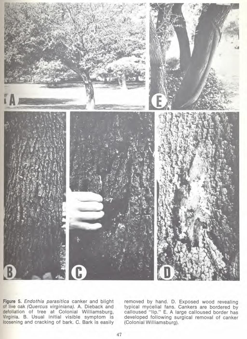
53
54 LITERATURE CITED Alexander, T. R PLANT SUCCESSION ON KEY LARGO, FLORIDA, INVOLVING PINUS CARIBAEA AND QUERCUS VIR- GINIANA. Quart. Jour. Fla. Acad. Sci. 16: Anderson, P. J. and H. W. Anderson THE CHESTNUT BLIGHT FUNGUS AND A RE- LATED SAPROPHYTE. Penna. Chestnut Tree Blight Comm. Bull. 4:1-26. Anderson, P. J. and W. H. Rankin ENDOTHIA CANKER OF CHESTNUT. N. Y. (Cornell) Agr. Exp. Sta. Bull p Anon WORK CONFERENCE REPORT ON CANKER OF LIVE OAK. Unpublished report of work conference of federal and state plant pathologists held at Atlanta, GA, May 18, p. Appel, D. N BIOLOGY AND PHYSIOLOGY OF PIN OAK BLIGHT CAUSED BY ENDOTHIA GYROSA. Ph.D. Dissertation Research, Unpublished. Batson, W. E. and W. Witcher LIVE OAK CANKERS CAUSED BY ENDOTHIA PARASITICA. Phytopathology 58: Bazzigher, G BEITRAG ZUR KENNTNIS DER ENDOTHIA PARA- SITICA ( MURR.) AND., DEM ERREGER DES KASTAN- IENSTERBENS. Phytopath. Z. 21: Benko, M. and J. Wimberley LANDSCAPE GROUP MEETS IN ILLINOIS. Amer. Nurseryman 131:9, 114, 115. Biraghi, A NUOVI OSPITI DI ENDOTHIA PARASITICA. Ann. Sper. Agr. N.S. 4: Boerboom, J. H. A. and P. W. Th. Maas CANKER OF EUCALYPTUS GRANDIS AND E. SALIGNA IN SURINAM CAUSED BY ENDOTHIA HA- VANENSIS. Turrialba 20: Bruner, S. C A NEW SPECIES OFENDOTHIA. Mycologia 8: Chona, B. L. and R. L. Munjal NOTES ON MISCELLANEOUS INDIAN FUNGI. I. Indian Phytopathology 3: Clapper, R. B., G. F. Gravatt, and D. C. Stout ENDOTHIA CANKER ON POST OAK. Plant Dis. Rep. 30: 381. Clinton, G. P CHESTNUT BARK DISEASE, ENDOTHIA GYROSA VAR. PARASITICA ( MURR.) CLINT. In Conn. Agr. Exp. Sta. Ann. Rpt. 1911/12. p Collingwood, G. H. and W. D. Brush KNOWING YOUR TREES. American Forestry Assoc., Washington, D. C. 349 p. Cooke, M. C SYNOPSIS PYRENOMYCETEN. Grevillea (London) 13: Darpoux, H CHESTNUT CANKER CAUSED BY ENDOTHIA PARASITICA. Rev. Tech. Prot. Veg. I, 4, p Darpoux, H LE CHANCRE DU CHATAIGNER CAUSE PAR ENDOTHIA PARASITICA. Institut national de la recherche agronomique. Document phytosanitaire 7. Dunlap, A. A. and A. L. Harrison DYING OF LIVE OAKS IN TEXAS. Phytopathology 39: Earle, F. S SOME FUNGI FROM PUERTO RICO. Muehlenbergia 1: Emert, G. H., R. J. Stipes, R. D. Brown, Jr., and J. E. Talmadge DIFFERENTIATION OF ENDOTHIA GYROSA AND E. PARASITICA BY DISC GEL ELECTROPHORESIS. Phytopathology 63:801. ( Abstr.) Fowells, H. A SILVICS OF FOREST TREES OF THE UNITED STATES. USDA Forest Service Agric. Hdbk p. Fries, D. M SUMMA VEGETABILIUM. Scandivaviae sect. 2. p Upsaliae. Fuckel, L EXSICCATI RHENANI HEDWIGIA 5:28. Fulton, H. R RECENT NOTES ON THE CHESTNUT BARK DIS- EASE. In Penna. Chestnut Blight Conf. Rpt p Gravatt, G. F THE CHESTNUT BLIGHT IN ASIA AND NORTH AMERICA. Unasylva 3:2-7. Gravatt, G. F BLIGHT ON CHESTNUT AND OAKS IN EUROPE IN Plant Dis. Rep. 36: Graves, A. H RELATIVE BLIGHT RESISTANCE IN SPECIES AND HYBRIDS OF CASTANEA. Phytopathology 40: Gruenhagen, R. H LIVE OAK DECLINE IN VIRGINIA. Plant Dis. Rep. 49:269. Halliwell, R. S LIVE OAK DECLINE. Proc. 40th Intl. Shade Tree Conf., Houston, Texas. p Halliwell, R. S ASSOCIATION OF CEPHALOSPORIUM WITH A DECLINE OF OAK IN TEXAS. Plant Dis. Rep. 50: Ham, D. L THE ETIOLOGY OF ENDOTHIA PARASITICA ON SCARLET OAK. M. S. Thesis, Duke University, Durham, N.C. 25p. Headland, J. K., G. J. Griffin, R. J. Stipes, and J. R. Elkins SEVERITY OF NATURAL ENDOTHIA PARASITICA INFECTION OF CHINESE CHESTNUT. Plant Dis. Rep. 60: Heald, F. D INTRODUCTION TO PLANT PATHOLOGY, 2ND ED. McGraw-Hill Book Company, New York. 603 p. Hepting, G. H DISEASES OF FOREST AND SHADE TREES OF THE UNITED STATES. USDA Forest Service Agric. Hdbk. No p. Hunter, P. P THE BLIGHT AND CANKER OF PIN OAK ( QUERCUS PALUSTRIS MUENCHH.) INCITED BY ENDOTHIA GYROSA ( SCHW.) FR.: SOME FACTORS AFFECTING DISEASE DEVELOPMENT. Ph. D. Dissertation, Va. Polytech. Inst. & State Univ. Library, Blacksburg. 113 p. Kobayashi, T NOTES ON THE GENUS ENDOTHIA IN JAPAN. II. A NEW SPECIES HAVING FOUR-SPORED ASCI. Trans. Mycol. Soc. Japan 6: Kobayashi, T TAXONOMIC STUDIES OF JAPANESE DIAPORTH- ACEAE WITH SPECIAL REFERENCE TO THEIR LIFE HISTORIES. Bull. Govt. Forest Expt. Sta p. Tokyo, Japan. Kobayashi, T. and K. Ito TWO NEW SPECIES OF ENDOTHIA. Ann. Phytopath. ii Soc. Japan 21:
55 Lindgren, R. M., R. P. True, and E. R. Toole SHADE TREES FOR THE SOUTHEAST. In Trees, Yearbook of Agriculture, U. S. Dept. of Agric., Washington, D. C. 944 p. Little, E. L., Jr CHECK LIST OF THE NATIVE AND NATURALIZED TREES OF THE UNITED STATES INCLUDING ALASKA. USDA Forest Service, Washington, D. C. 325 p. May, C. and R. W. Davidson ENDOTHIA PARASITICA ASSOCIATED WITH A CANKER OF LIVE OAK. Plant Dis. Rep. 44: 754. May, C SHADE TREES FOR THE HOME. USDA Agric. Hdbk. No p. Merkel, H. W A DEADLY FUNGUS ON THE AMERICAN CHEST- NUT. In 10th Ann. Rept. N. Y. Zool. Soc p Muller, E. and J. A. von Arx DIE GATTUNGEN DER DIDYMOSPOREN PYRENO- MYCETEN. Beitr. zur. Kryptogamenfl. Schweiz. Band 11, Heft p. Negru, A. and S. Mozes NEW AND RARE FUNGI FOUND ON PEAT BOGS "MOHOS" IN THE PEOPLE'S REPUBLIC RUMANIA. Akad. Nauk. Armianskoi SSR Verevan. Isvestiia. Biologicheskia Nauki Gerevan 18: Nutman, F. J. and F. M. Roberts ACUTE DIE-BACK OF CLOVE TREES IN THE ZANZIBAR PROTECTORATE. Ann. Appl. Biol. 39: Otth, G SECHSTER NACHTRAG ZU DEM IN NR DER MITTHEILUNGEN ENTHALTENEN VERZEICHNISSE SCHWEIZERISCHER PILZE. Naturforschende Gesellschaft, Bern. Mitteilungen. p Peacher, P. H LIVE OAK IN MISSISSIPPI INFECTED BY EN- DOTHIA PARASITICA. Plant Dis. Rep. 53:304. Phelps, W. R LIVE OAK IN NORTH CAROLINA AND FLORIDA INFECTED BY ENDOTHIA PARASITICA. Plant Dis. Rep. 58: Pirone, P. P TREE MAINTENANCE. 4th ed. Oxford Univ. Press, N. Y. 574p. Reid, J. and C. Booth SOME SPECIES SEGREGATED FROM THE GENERA CRYPTOSPORA, CRYPTOSPORELLA AND SILLIA. Can. J. Bot. 47: Roane, M. K., R. J. Stipes, P. M. Phipps, and 0. K. Miller, Jr ENDOTHIA GYROSA, CAUSAL PATHOGEN OF PIN OAK BLIGHT. Mycologia 66: Roane, M. K. and R. J. Stipes THE GENUS ENDOTHIA: A REAPPRAISAL. Va. J. Sci. 25: 74 (Abstr.). Roane, M. K., R. J. Stipes, and J. R. Elkins PIGMENT ANALYSIS OF FRUITING STRUCTURES AND MYCELIUM IN THE GENUS ENDOTHIA. Va. J. Sci. 26:65 ( Abstr. ). Roane, M. K. and R. J. Stipes THE FUNGAL GENUS ENDOTHIA IN THE SOUTH- ERN APPALACHIANS. In Distributional History of the Biota of the Southern Appalachians, Part IV: Algae and Fungi. Univ. Press of Va., Charlottesville. p Seymour, A. B HOST INDEX OF THE FUNGI OF NORTH AMERICA. Harvard Univ. Press, Cambridge, Mass. 732 p. Shear, C. L., N. E. Stevens, and R. J. Tiller ENDOTHIA PARASITICA AND RELATED SPECIES. U. S. Dept. Agr. Bull p. Snow, G. A., J. S. Beland, and F. J. Czabator FORMOSAN SWEETGUM SUSCEPTIBLE TO NORTH AMERICAN ENDOTHIA GYROSA. Phytopathology 64: Stipes, R. J. and T. C. Davis DISEASES OF LANDSCAPE TREES OBSERVED IN VIRGINIA. Plant Dis. Rep. 56: Stipes, R. J., P. P. Hunter, J. R. Elkins, and M. K. Roane ENDOTHIA CANKERS OF OAKS. In Proc. First Metropolitan Tree Improvement Alliance Conference, Lanham, MD., July, (USDA Forest Service, Broomall, PA, In Press(. Stipes, R. J. and P. M. Phipps. 1971a. A SPECIES OF ENDOTHIA ASSOCIATED WITH A CANKER DISEASE OF PIN OAK (QUERCUS PALUSTRIS) IN VIRGINIA. Plant Dis. Rep. 55: Stipes, R. J. and P. M. Phipps. 1971b. CURRENT STATUS OF ENDOTHIA CANKER OF LIVE OAK IN VIRGINIA. Plant Dis. Rep. 55:201. Stipes, R. J., P.M. Phipps, and 0. K. Miller, Jr A PIN OAK BLIGHT ASSOCIATED WITH A SPECIES OF ENDOTHIA. Va. J. Sci. 22:86 (Abstr.). Stipes, R. J. and J. L. Ratliff EFFECT OF TEMPERATURE ON LINEAR GROWTH OFENDOTHIA GYROSA. Va. J. Sci. 24:136 (Abstr.). Stipes, R. J. and M. K. Roane EFFECTS OF ph AND TEMPERATURE ON THE GROWTH OF TEN ENDOTHIA SPECIES. Va. J. Sci. 27:60 ( Abstr.). Taubenhaus, J. J LIVE OAK DISEASE IN AUSTIN. 47th Ann. Rep., Texas Agr. Exp. Sta. Taubenhaus, J. J LIVE OAK DISEASE IN AUSTIN. 48th Ann. Rept., Texas Agr. Exp. Sta. Theissen, R. and H. Sydow DIE DOTHIDEALES. Annales Mycologici 13: Van Arsdel, E. P SOME CANKERS ON OAKS IN TEXAS. Plant Dis. Rep. 56: Van Arsdel, E. P. and R. S. Halliwell PROGRESS IN RESEARCH ON LIVE OAK DECLINE. Plant Dis. Rep. 54: Vizioli, J SOME PYRENOMYCETES OF BERMUDA. Mycologia 15: Wehmeyer, L. E CULTURAL STUDIES OF THREE NEW PYRENO- MYCETES. Mycologia 28:35-46.
56 An Evaluation of the Fungal Genus Endothia Martha K. Roane and R. J. Stipes Department of Plant Pathology and Physiology, Virginia Polytechnic Institute and State University, Blacksburg, VA ABSTRACT. Of the several species ascribed to the genus Endothia or its imperfect state, Endothiella, four, Endothia sordida, Endothia parryi, Endothia nitschkei and Endothiella robiniae, have been removed from the genus. Since materials of Endothia tetraspora and Endothiella simoniani have not been examined, the status of these taxa is uncertain. The synonymy of E. longirostris with E. radicalis and of E. tropicalis with E. havanensis is being studied. The position of E. eugeniae is doubtful because of the atypical stromata, the absence of bisanthraquinone pigments and the ovoid rather than bacilliod conidia. Endothia coccolobii, E. gyrosa, E. havanensis, E. japonica, E. macrospora, E. parasitica, E. radicalis, E. singularis, and E. viridistroma are retained as discrete species. Endothia Fries The genus Endothia, described by Fries (1849) from specimens of Sphaeria gyrosa Schw. ( Schweinitz, 1822), and its imperfect state, Endothiella Sacc. ( Saccardo, 1906), today contain 18 species aside from the synonyms listed in Shear et al. (1917). Endothia is characterized by stromata subcortical in origin, variable in size and shape, pustular to subspherical, subcoriaceous to friable, sometimes confluent, surface light auburn or chestnut to mahogany red (dark olive green in one species), capucine yellow or cadmium orange to scarlet within ( dark olive green in one species); pycnidial and perithecial stromata the same or similar; pycnidia few to numerous, consisting of simple cavities or complex and irregular chambers; conidia minute, simple, bacilliform to oblong, yellowish to reddish in mass; perithecia deeply immersed, in one or more irregular layers, usually black when mature, with long necks, black within, colored like the stroma without; asci clavate to oblong-fusoid, 8-spored, usually without paraphyses; ascospores oblongfusoid or subellipsoid to cylindric or allantoidcylindric, one-celled or two-celled, hyaline to pale yellow. There are two sections in the genus, the first of which is made up of species having one-celled ascospores and the second having species with twocelled ascospores. Section 1. Ascospores one-celled Endothia gyrosa (Schw.) Fr. As we know the type of the genus today, it is characterized by bright orange to chestnut pulvinate stromata, mm diameter by mm high; one-celled, oblong, hyaline conidia, 3-4 x pm, formed in labyrinthiform chambers in the stromata and exuded in droplets; dark brown to black membranous, slender-necked perithecia, pm diameter, embedded in the stromata; oblong-fusoid or subclavate asci, x 6-7 pm, containing eight cylindric to allantoid, one-celled, hyaline ascospores, 7-11 x 2-3,um. Endothia singularis (H. & P. Syd.) Shear & Stevens The second member of the section was described as Calopactis singularis by H. & P. Sydow (1912) and was removed to Endothia in Shear et al. (1917). Characteristics of this species are the very large (3-5 mm diameter by 2-4 mm high) scarlet stromata; one-celled oblong hyaline conidia, 6-8 x 1 pm, formed in nearly spherical cavities, pm diameter, embedded in the stromata and set free in a powdery mass by the disintegration of the stroma wall; dark brown to black membranous, slender-necked perithecia, pm diameter, embedded in the stromata; oblong-cylindric to fusoid or subclavate asci, x pm, containing eight cylindric to allantoid, hyaline, one-celled ascospores, 6-11 x pm. Endothia viridistroma Weh. The third member of the section, described by Wehmeyer (1936) has dark olive-green tuberculate stromata, 2-5 mm diameter by mm high; onecelled, hyaline, cylindric to allantoid conidia, x pm, formed in irregular locules surrounded by a greenish-black pseudo-parenchymatous wall in the stromata and set free by formation of yellow spore tendrils; dark brown to black membranous, slender-necked perithecia, x pm, embedded in the stromata; clavate asci, x 3-4 pm, containing eight allantoid, hyaline, one-celled ascospores, 5-6 x pm. Endothia tetraspora Kobayashi Material of the last member of the section described by Kobayashi (1965) was not available for study. As Kobayashi (1970) noted, it has an atypical, small, compact, yellow-orange ectostroma and scanty entostroma; no conidia were reported; dark brown to black walled perithecia, µ m diameter, embedded beneath the disc-like ectostroma, with confluent black necks erumpent through the stroma as in Valsa; clavate to oblong-clavate asci, x pm, containing four one-celled, hy-
57 aline, fusoid or elliptic ascospores, x Am. Kobayashi found some characteristics common to this species and certain members of Cryptosporella. Further work is necessary to determine whether this species belongs in Endothia or in another genus. Section 2. Ascospores two-celled Endothia radicalis (Schw.) Ces. & DeNot. Endothia fluens ( Sow. ) Shear and Stevens is properly placed in synonymy with E. radicalis since the former was based upon Sphaeria fluens Sow. described by Sowerby (1814) in its pycnidial condition. The name E. fluens was quite correct for Shear et al. (1917) since they were observing the American Rules of Nomenclature. However, under the International Rules of Nomenclature now being observed, the name first applied to the sexual state of a fungal taxon takes priority. Therefore, the name E. radicalis (Schw.) Ces. & DeNot. takes precedence since it is based on the description of an ascosporic specimen. This species was first described as Sphaeria radicalis by Schweinitz ( 1832 ) and was removed to Endothia by Cesati and DeNotaris (1863). It is characterized by pulvinate, light auburn to chestnut stromata, mm diameter by mm high, containing irregular pycnidial chambers with oblong, hyaline, one-celled conidia, 3-5 x 1.5 x 2µm; dark brown to black perithecia, Am diameter, with long slender black necks, containing oblong-fusoid or subclavate asci, x 6-8 mm, with eight hyaline, 2-celled, oblong-fusoid or subellipsoid ascospores, not constricted at the septum, 6-10 x Am. Endothia sordida Fuckel The material of this species described by Fuckel (1866) that we examined (E. sordida Fuckel. Fungi Rhenani #1586. Type. National Fungus Collection) had stromata that were sandy brown to dark brown on the outside, chocolate brown within; black necks of perithecia forming papillae on the surface of the stromata, erumpent through the bark and scarcely distinguishable from the color of the bark, asci 8-spored, cylindric, 118 x 13 mm; 2-celled, brown, ellipsoid ascospores, 20 x 10 Am, with conspicuous globules. Because of the color of the stromata and the quite large, ellipsoid, brown ascospores with conspicuous globules, this species is removed from the genus Endothia. Endothia nitschkei Otth Otth (1868) in describing this species said that the base of the tawny-rusty brown tubercle-like stroma was covered with dark brown to black membranous sacs of hyaline, cylindric, one-celled conidia, 5 x 1 pm, that were expelled as a coarse white tendril. The perithecia were immersed in the bark with erect, converging necks piercing through the stroma with few black exerted ostioles. The 2-celled, oblong, blunt, faintly colored ascospores, x 4-5 mm, were constricted at the septum. When the perithecia mature in the spring following the fall maturation of the conidia, the stromata become whitish due to weathering on the outer surface. We were unable to find stromal tissue near the perithecia which we examined in material from the National Fungus Collection (E. nitschkei Otth. Herbier Fuckel #739). No pigments from tissue of this specimen dissolved in absolute ethanol in contrast to the almost immediate solution of pigments of E. gyrosa, E. radicalis, etc. Because of the perithecia immersed in the bark rather than in stromata, with converging necks like Valsa, and the absence of pigments soluble in absolute ethanol, this species is removed from the genus Endothia. Endothia parryi Earl. The description of this species is included by Cooke (1885) in his Synopsis Pyrenomyceten. However, Theissen and Sydow (1915) removed the taxon to the genus Dothidella. We concur with this disposition after examination of specimens from the National Fungus Collection and the Farlow Herbarium. Endothia longirostris Earle Characteristics of the species described by Earle ( 1901) are the yellow to chestnut stromata, 1-3 mm diameter; with oblong-elliptic, hyaline, one-celled conidia 2-4 x Am, produced in labyrinthiform cavities and expelled from a single large pore or irregular rupture at the apex of the stroma in a stout coral-red spore tendril; black, membranous, longnecked, up to 1 cm perithecia, m m diameter, embedded in the stromata usually at the base of the pycnidial locules; oblong-cylindric to fusiform asci, x 5-7µm, with eight 2-celled, hyaline, ovoid to ovoid-elliptical ascospores, x 3-4 Am. According to Kobayashi ( 1970), E. longirostris is a synonym of E. radicalis. However, we (1978) have found that stromal tissue of E. longirostris contains the bisanthraquinones, skyrin and rugulosin, while E. radicalis contains only skyrin. Also, in numerical analysis of percent similarity of Endothia spp., we (1976) found that E. radicalis (=E. fluens) and E. longirostris were 28 percent similar, a value that does not indicate a very close relationship. Until we obtain fresh material of both, we prefer to retain E. radicalis and E. longirostris as separate species. Endothia parasitica ( Murr.) P. J. & H. W. And. The fungus found associated with a serious canker of the American chestnut in the New York Zoological Park was described as Diaporthe parasitica ( Murrill, 1906) and Endothia gyrosa var. parasitica ( Clinton, 1912). It was removed from Diaporthe and placed in Endothia as E. parasitica by P. J. and H. W. Anderson ( 1912). However, in the early reports of work on the chestnut blight organism there is much confusion among E. gyrosa, E. radicalis (=E. fluens) and E. parasitica because of the close resemblance of the asexual states of these species ( Roane and Stipes, 1978). Ascosporic voucher specimens remain the best way of separat-
58 ing these species at present, although certain cultural characteristics, pigment analysis and protein and enzyme analysis are also valuable. Endothia parasitica is characterized by yellow to auburn, corticular, slightly erumpent to truncate conical, usually separate but frequently confluent stromata, mm diameter by mm high; hyaline, oblong to cylindric, one-celled conidia, 3-5 x pm, formed in irregular cavities in the stromal tissue and expelled in spore tendrils that are yellow when fresh and coral red when old; dark brown to black, membranous, globose to flask-shaped, blacknecked perithecia, Am diameter, embedded in the stromata; oblong-elliptical to subclavate asci, x 7-9 Am, containing eight hyaline, ellipsoid, 2- celled ascospores, 7-11 x pm, sometimes constricted at the septum, with a gelatinous envelope. In the cambium and bark of the host this fungus typically produced yellow or buff fan-shaped formations of mycelium. These are not produced by E. gyrosa or E. radicalis. Endothia havanensis Bruner In his description of this fungus Bruner (1916) compared it with cultures and specimens of E. longirostris and E. radicalis and concluded that the three represented different species. Endothia havanensis is denoted by rounded, wart-shaped to subelongate, bright-yellow to orange to yellowbrown stromata, 2-15 by 2-4 mm, containing irregular pycnidial chambers with hyaline, rodshaped, one-celled, conidia, 3-4 x pm, discharged in yellow or orange-yellow spore tendrils; black, globlose, long-necked perithecia, A m diameter, deeply embedded in stromata; subclavate, 8-spored asci, x Am, with hyaline, fusoid 2-celled ascospores, x Am, constricted at the septum. Endothia tropicalis (Berk. & Br.) Shear & Stevens In 1875, Berkeley and Broome described this fungus and named it Diatrype gyrosa. Shear and Stevens ( Shear et al., 1917) studied material collected by Petch in Ceylon in 1913 and removed it from Diatrype to Endothia but had to supply a new specific epithet, tropicalis, since gyrosa was preempted by the earlier E. gyrosa (Schw. ) Fr. Endothia tropicalis has orange chrome to sanford brown, pustular to pulvinate stromata, 1-5 mm diameter, with numerous irregular cavities containing the hyaline, oblong to cylindric, one-celled conidia, x Am ; black, membranous, slender-necked, globose perithecia, µ m diameter, with oblong or subclavate, 8-spored asci containing 2-celled, not constricted at the septum, hyaline, subelliptical ascospores, x Am, with a gelatinous envelope. Endothia coccolobii Vizioli A fungus on sea grape collected by H. H. Whetzel in Bermuda was described and named E. coccolobii by Vizioli (1923), who concluded that E. longirostris was closely related to but not the same as this species. Its characters are small, orange-rufous, hemispheric to conoid, erumpent stromata, mm diameter, containing irregular pycnidial locules with hyaline, bacilliform conidia, 2-3 x Am, discharged through a single black conoid papilla; leathery-membranous, coffee-black, long slendernecked, globose to subglobose perithecia, Am diameter, containing oblong to subclavate 8-spored asci, x 4-6 Am, with hyaline, 2-celled, not constricted at the septum, ovoid to fusoid ascospores, x Am. Endothia japonica Kobayashi & Ito Endothia japonica Kobayashi & Ito (1956) was placed in synonymy with Otth's E. nitschkei by Kobayashi (1970). Since we have removed E. nitschkei sensu Otth from Endothia, we retain the original name of this species of Kobayashi & Ito. It is characterized by yellow-orange or brown-orange, erumpent, conic to truncate-conic stromata, 2-3 mm diameter by 1-2 mm high; dark brown to black, globose, long-necked perithecia, pm diameter, with clavate to oblong-clavate, 8-spored asci, x pm, elliptic, 2-celled, constricted at the septum, hyaline to pale brown ascospores, x Am ; conidial stromata smaller and paler than perithecial stromata, with multilocular cavities containing hyaline, one-celled, allantoid or rod-shaped conidia, 3-4 x Am, discharged as sticky yellow tendrils. Endothia macrospora Kobayashi & Ito This fungus, described by Kobayashi & Ito (1956), typically has orange, erumpent, pulvinate stromata, 1-3 mm diameter by 1-2 mm high, containing irregular pycnidial locules with hyaline, one-celled, allantoid or rod-shaped conidia, x /I m, discharged through a central pore as sticky yellow tendrils; dark brown membranous, globular, longnecked perithecia, pm diameter, embedded in lower part of stroma, containing clavate to oblong-clavate, 8-spored asci, x pm, with elliptic, 2-celled, slightly constricted at the septum, hyaline ascospores, x Am. As inferred by the specific epithet, macrospora, this species has the largest ascospores of this section of the genus. Endothia eugeniae (Nutman & Roberts) Reid & Booth The causal agent of a die-back of clove was named Cryptosporella eugeniae by Nutman and Roberts (1952) and was later transferred to Endothia by Reid and Booth (1969). This species is typified by orange to rust brown, erumpent, conic stromata, mm diameter by 0.5 mm high, containing one to several irregular pycnidial locules with one-celled, oval, hyaline conidia, x Am, extruded in yellow tendrils; dark brown to black, globose, longnecked perithecia, pm diameter, containing clavate, 8-spored asci, x Am, with oblong to elliptical, 2-celled, not constricted at the septum,
59 hyaline ascospores, x 2-3 Aim, widest in the upper cell. Nutman and Roberts described the stromata as valsiform and noted that perithecia contained in the same stroma have converging necks. Reid and Booth considered this taxon typical of the genus Endothia because of the nature of the stroma, the nature of the pycnidia, the orange-brown coloration of the perithecial necks and papillae, the arrangement of the perithecia within the stroma, and the possession of two-celled ascospores. However, in our study of specimens of other species in the genus, we have found that these species have much more abundant stromal tissue that contains at least one bisanthraquinone ( Roane and Stipes, 1978) and have not found this to be true of E. eugeniae. Cylindric or bacilliform conidia are typical of all members of the genus except this one which has oval conidia. Because of these deviations from the normal pattern in Endothia we regard this species as a doubtful member of the genus Endothia. Endothiella Saccardo. Imperfect state of Endothia Endothiella robiniae Chona & Munjal The slate-black, erumpent, pulvinate, stalked stromata, mm diameter, are dark brown within and produce conidiophores and conidia on the outer surface rather than in pycnidial locules submerged in the stromal tissue ( Chona & Munjal, 1950). The hyaline, cylindric-fusoid to allantoid, one-celled, very variable conidia, 8µm or more long, with papillae at both ends, are born on very long conidiophores, m. When we placed stromal tissue of this taxon in lactophenol, no yellow to orange coloration appeared. We find from our examination of the fungus ( Specimen 19759, Herb. Crypt. Ind. Orient. Fungi Indian Agricultural Research Institute, New Delhi) that it is not a species of Endothiella because of the black, stalked stroma with an external conidial layer, the atypical characters of the conidia and conidiophores, and the absence of the yellow and orange bisanthraquinone stromal pigments. It is probably the imperfect state of a species of Hypoxylon. Endothiella simoniani Negru and Mozes We reserve judgment on this fungus described by Negru and Mozes (1965) since we have not seen it, although from the Latin description it seems to have characters similar to those of Endothiella. Negru and Mozes did not compare it with other members of the genus. DISCUSSION In 1951, Orsenigo divided Endothia into two subgenera, the first of which he called Proendothia and the second, Euendothia. He placed E. gyrosa and E. singularis in subgenus Proendothia and E. radicalis, E. longirostris, and E. tropicalis in subgenus Euendothia. He regarded E. radicalis as made up of two subspecies, ssp. aflabellata (=E. fluens [ Sow.] Shear and Stevens) and ssp. parasitica (=E. parasitica [ Murr.] P. J. and H. W. And.). However, we (Roane and Stipes, 1978; Stipes et al., 1978) have found that E. parasitica and E. radicalis can be differentiated by both pigment analysis and enzyme analysis and with the addition of these two characters to others previously known we consider these to be distinct species. Whether or not Endothia should be subdivided into the two subgenera as proposed by Orsenigo, divided into two genera, or left intact has not been considered by us at this time. Kobayashi (1970) proposed that E. fluens and E. longirostris be placed in synonymy with E. radicalis, that E. japonica be placed in synonymy with E. nitschkei and that E. tropicalis be placed in synonymy with E. havanensis. We have already discussed the status of the first two synonymies. However, the situation regarding E. tropicalis and E. havanensis needs further clarification since, at present, there is insufficient material of E. havanensis available for comparison with E. tropicalis. Two aids for the identification of an isolate obtained from a specimen containing only conidial stromata and presumed to be E. gyrosa, E. parasitica, or E. radicalis are 1) checking for the production of perilla purple by the culture when grown on white cornmeal medium and 2) analyzing the bisanthraquinone content by thin-layer chromatography. Endothia gyrosa and E. parasitica contain skyrin, oxyskyrin and rugulosin while E. radicalis contains only skyrin. Both E. gyrosa and E. radicalis produce perilla purple on white cornmeal but E. parasitica does not. In addition, we ( Stipes et al., 1978) have found that these three species have different electrophoretic enzyme and protein patterns. We conclude that the genus Endothia contains the following species: E. coccolobii, E. gyrosa, E. havanensis, E. macrospora, E. parasitica, E. radicalis, E. singularis and E. viridistroma. We prefer not to consider E. longirostris as a synonym of E. radicalis and wish to obtain for study more fresh material of both species before making a final judgment. The same situation applies to the case of E. havanensis and E. tropicalis. As previously stated, we conclude that the original E. nitschkei Fuckel should be removed from the genus. Therefore, Kobayashi's species originally described as E. japonica is removed from synonymy with E. nitschkei and retains its first name. We reserve judgment on the status of E. eugeniae until further material can be obtained for study. The status of Endothia tetraspora and Endothiella simoniani is uncertain since we were unable to examine specimens of these species. ACKNOWLEDGMENTS The assistance of R. W. Davidson, J. Dingley, F. Hawksworth, T. Kobayashi, G. Samuels, Centraalbureau voor Schimmelcultures, Commonwealth
60 Mycological Institute, Cornell University Plant Pathology Herbarium, Farlow Herbarium, Indian Agricultural Research Institute Herbarium, National Fungus Collection, New York Botanical Garden, National Mycological Herbarium-Ottawa in obtaining cultures and specimens is gratefully acknowledged. LITERATURE CITED Anderson, P. J. and H. W. Anderson THE CHESTNUT BLIGHT FUNGUS AND A RE- LATED SAPROPHYTE. Phytopathology 2: Berkeley, M. J. and C. E. Broome ENUMERATION OF THE FUNGI OF CEYLON. J. Linn. Soc. (London) Bot. 15: Bruner, S. C A NEW SPECIES OF ENDOTHIA. Mycologia 8: Cesati, Vincenzo and G. denotaris SCHEMA DI CLASSIFICAZIONI DEGLI SFERIACEI ITALICI ASCHIGERI. In Comm. Soc. Crittog. Ital. 1:240. Chona, B. L. and R. Munjal NOTES ON MISCELLANEOUS INDIAN FUNGI. I. Indian Phytopathology III: Clinton, G. P THE RELATIONSHIPS OF THE CHESTNUT BLIGHT FUNGUS. Science, n. s. 36: Cooke, M. C SYNOPSIS PYRENOMYCETEN. Grevillea. London 13: Earle, F. A SOME FUNGI FROM PORTO RICO. Muhlenbergia 1: Fries, E. M SUMMA VEGETABILIUM. Scandinaviae Sect. 2. p Upsaliae. Fuckel, K. W. G. L EXSICCATI RHENANI. Hedwigia 5:28. Kobayashi, Takao NOTES ON THE GENUS ENDOTHIA IN JAPAN II. A new species having four-spored asci. Trans. Myc. Soc. Jap. 6: Kobayashi, Takao TAXONOMIC STUDIES OF JAPANESE DIAPOR- THACEAE WITH SPECIAL REFERENCE TO THEIR LIFE- HISTORIES. Bull. Govt. For. Expt. Sta Tokyo, Japan. 242 p. Kobayashi, T. and K. Ito TWO NEW SPECIES OF ENDOTHIA. Ann. Phytopath. Soc. Japan 21: Murrill, W. A A NEW CHESTNUT DISEASE. Torreya 6: Negru, A. and S. Mozes NEW AND RARE FUNGI FOUND ON PEAT BOGS "MOHOS" IN THE PEOPLE'S REPUBLIC RUMANIA. Akademia Nauk. Armianskoi SSR Verevan. Izvestiia. Biologicheskia Nauki yerevan. 18: Nutman, F. J. and F. M. Roberts ACUTE DIE-BACK OF CLOVE TREES IN THE ZAN- ZIBAR PROTECTORATE. Ann. Appl. Biol. 39: Orsenigo, M CONTRIBUTO ALLA CONOSCENZA DEL GENERE ENDOTHIA I. ANALISE STATISTICA IN DELLE DIMEN- SION' DELLE ASCOSPORE DI ALCUNE SPECIE. II. IDENTIFICATION FRA E. PARASITICA ( Murr.) P. J. et H. W. And. ed E. fluens (Sow.) S. et S. e Toro fusione in E. radicalis (Schw.) DeNot. Considerazione sulla patogenecita delle specie e sulla classificazione del genere. Phytopath. Z. 18(2 ): Otth, G SECHSTER NACHTRAG. SU DEM IN NR DER MITTHEILUNGEN ENTHALTENEN VERZEICHNISSE SCHWEIZERISCHER PILZE. Naturforschende Gesellschaft, Bern. Mitteilungen. pp Reid, J. and C. Booth SOME SPECIES SEGREGATED FROM THE GENERA CRYPTOSPORA, CRYPTOSPORELLA, AND SILLIA. Can. J. Bot. 47: Roane, Martha K. and R. J. Stipes THE FUNGAL GENUS ENDOTHIA IN THE SOUTH- ERN APPALACHIANS. In: Distributional History of the Biota of the Southern Appalachians. Part IV: Algae and Fungi. Univ. Press of Virginia, Charlottesville. pp Roane, Martha K. and R. J. Stipes PIGMENTS IN THE FUNGAL GENUS ENDOTHIA. Va. J. Sci. 29. Saccardo, P. A NOTAE MYCOLOGICAE, SERIES VII. Ann. Mycol. 4 : Schweinitz, L. D. Von SYNOPSIS FUNGORUM CAROLINAE SUPERIOR'. Leipzig. 105 pp. Schweinitz, L. D. Von SYNOPOSIS FUNGORUM IN AMERICAN BOREAL' MEDIA DEGENTIUM In: Trans. Amer. Phil. Soc., n.s. 4: Shear, C. L., N. E. Stevens, and R. J. Tiller ENDOTHIA PARASITICA AND RELATED SPECIES. USDA Bull. 380, 82 pp. Sowerby, James COLORED FIGURES OF ENGLISH FUNGI OR MUSHROOMS. Sup. London. Stipes, R. J., G. H. Emert, R. D. Brown, Jr., and M. K. Roane DIFFERENTIATION OF ENDOTHIA GYROSA, E. PARASITICA AND E. RADICALIS BY DISK ELECTRO- PHORESIS OR ULTRAMYCELIA ENZYMES AND PRO- TEINS. Va. J. Sci. 29. Sydow, H. and P. Sydow NOVAE FUNGORUM SPECIES - VII. Ann. Mycol. 10: Theissen, F. and H. Sydow DIE DOTHIDEALES. Ann. Mycol. 13: Vizioli, J SOME PYRENOMYCETES OF BERMUDA. Mycologia 15: Wehmeyer, L. E CULTURAL STUDIES OF THREE NEW PYRENOMY- CETES. Mycologia 28:35-46.

61 Virulence of Endothia parasitica Isolated from Surviving American Chestnut Trees Gary J. Griffin Virginia Polytechnic Institute and State University John R. Elkins Bluefield State College, Bluefield, WV Gail Tomimatsu and Frederick V. Hebard Virginia Polytechnic Institute and State University, Blacksburg, VA ABSTRACT. Large surviving American chestnut trees may possess blight resistance if they are not infected with American hypovirulent strains of Endothia parasitica. To investigate this, isolates of E. parasitica were obtained in 1975, 1976 and 1977 from cankered tissues of large, surviving and small American chestnut trees ( Castanea dentata), and from cankered Chinese chestnut trees ( C. mollissima) growing in the Appalachian region. These tissue isolates and single-conidium progeny of tissue isolates were examined for pigment production, linear growth rate in vitro, tannin utilization and pathogenicity on disease-free American chestnut stump sprouts. A large variation in pathogenicity among isolates was observed. Most isolates from surviving American chestnut trees were moderately or highly pathogenic, when comparisons were made to two isolates from blight-killed American chestnut trees and a European-derived hypovirulent isolate, Ep43. Some isolates from a large American chestnut tree in Virginia were hypovirulent ( weakly pathogenic). INTRODUCTION The American chestnut ( Castanea dentata [Marsh.] Borkh. ) grew to be a large tree before Endothia parasitica ( Murr. ) P. J. & H. W. And. devastated the extensive natural stands in the Appalachian region. Today, only a few large trees (Fig. 1) remain in scattered locations throughout the region. Typically they are extensively infected with E. parasitica and show various degrees of dieback in the crown and bole due to this infection. American stump sprouts have become established since the blight but show similar or greater levels of disease. The residual trees may possess some degree of blight resistance if they are not infected with American hypovirulent strains of E. parasitica. This investigation was undertaken to examine this question for selected surviving American chestnut trees in Virginia, West Virginia, and other states within the natural range of the American chestnut. Figure 1. Surviving American chestnut tree in West Virginia from which Endothia parasitica was isolated for use in pathogenicity studies. METHODS AND MATERIALS In 1975, 1976 and 1977, bark samples were obtained at the canker margins of surviving American and Chinese chestnut (C. mollissima Bl. ) trees. Samples were placed in plastic bags before tissue pieces were transported to the laboratory and plated on acidified Difco potato dextrose agar ( APDA). Endothia parasitica isolates growing from these tissue pieces ( termed "tissue isolates") were trans-
62 ferred to PDA slants; mature cultures of these tissue isolates were stored, if necessary, under mineral oil until used in pathogenicity tests. Singleconidium isolates were obtained from mature tissue isolates soon after initial isolations were performed. Tests made on approximately 150 conidia of two representative isolates with Feulgen stain (Johansen, 1940) indicated that most conidia were uninucleate, but that some conidia (10-20 percent) contained two bodies, often close together, that stained. Degree of pigmentation and linear growth measurements of 20 single-conidium isolates, for each of several tissue isolates, were determined at room temperature (26-28 C) and in room light ( cool-white fluorescent) after 6 and 12 days incubation. Tissue isolates, together with the darkest pigmented and the lightest pigmented single-conidium isolates, were selected for pathogenicity trials. Most tissue isolates (W, AP, C, WE, SW, MC, IV, KE, RE, PL) were obtained from American chestnut trees with a diameter breast height of cm; other isolates ( D, AR, CH, CK, ALA) were obtained from American chestnut trees with a diameter breast height of cm. Isolates CCS, HF20 and HF38 were obtained from Chinese chestnut. European-derived ( Van Alfen et al., 1975) hypovirulent isolate, Ep43 ( obtained from R. A. Jaynes, Conn. Agr. Exp. Sta., New Haven), was used as a reference isolate in these trials. In addition, two E. parasitica isolates, CR and PC ( Table 1), from blight-killed American chestnut trees were used for reference. Blight-free American chestnut stump sprouts ( circa 2 to 4 cm diameter breast height) growing in the Jefferson National Forest near Blacksburg, Virginia, were used for pathogenicity tests. The cork-borer-agardisc method of inoculation was used. Five cork-borer wounds (0.7 cm diameter), approximately 50 cm apart, were made to the vascular cambium for each tree. Potato-dextrose-agar discs of five different E. parasitica isolates were inserted into the wounds on each tree, using aseptic technique. Inoculated wounds were covered with masking tape. Five replicate inoculations, on different trees, were made for each tissue isolate or single-conidium isolate. In 1976 and 1977, inoculations were made on June 1 and May 23, respectively; canker lengths were determined on August 14 and October 1 in 1976 and on August 3 and October 11 in Pathogenicity ratings of isolates were based on canker length measurements made at both time intervals. However, emphasis was placed on August measurements as some cankers were not measurable in October due to brown staining of the bark or to tree death. Utilization of American chestnut tannins by E. parasitica isolates was determined by inoculating 10 ml of aqueous bark extract (1 g powdered youngshoot bark/10 ml water) in a 20-ml screw-cap vial, with 10 6 conidia. Tannin assays were performed by the method of Elkins et al. (1977) on 0.5 ml subsamples removed from each of two vials per isolate after 17 days incubation at room temperature (21 C). RESULTS AND DISCUSSION Pigmentation and linear growth. All tissue isolates produced yellow-orange pigmented colonies on PDA, typical of E. parasitica ( Roane and Stipes, 1976; Shear et al., 1917), although there were often differences in colony texture, pigment intensity, pigment distribution in the colony and the time required for pigment production. Single-conidium progeny of almost all tissue isolates also varied to some degree in the intensity of pigmentation produced, as well as in the amount of linear growth produced after six days incubation (Table 1). Two tissue isolates ( D and AR), as well as some singleconidium progeny of these isolates ( Table 1), produced pigment at a much later stage in colony growth than other E. parasitica isolates. Unlike white Ep43 isolates, however, all D and AR isolates were pigmented after 12 days incubation in room light (Table 1). Both white and yellow-orange (normal) single-conidium progeny types were obtained from white Ep43 as has been found for white European hypovirulent strains ( Bonifacio and Turchetti, 1973; Grente and Sauret, 1969). However, in separate tests, light influenced the frequency of pigmented colonies and the rate of pigment formation by single-conidium progeny of white ( at six days) Ep43. Some tests were conducted at 25 C in a Sherer-Gillette Model CEL growth chamber in continuous darkness or continuous light (2,000 ft-c) supplied by cool-white fluorescent lamps. Other tests were conducted in the laboratory under room light. After six days of incubation, 14 of 23 dark-incubated monoconidial progeny on Difco PDA did not contain pigment, while only 1 of 23 light-incubated ( growth chamber) progeny did not contain pigment (were entirely white). Two of 20 room-light-incubated progeny were entirely white. After 12 days of incubation, 2 of 23 dark-incubated progeny were entirely white. In contrast, no light-incubated ( growth chamber) progeny were entirely white, and 1 of 20 room-lightincubated progeny was entirely white. At this time, light-incubated ( growth chamber) progeny exhibited greater intensities of yellow-orange pigment than progeny from the other two treatments. Although no information on pigmentation was given, Barnett and Lilly (1952) observed greater production of pycnidia by E. parasitica under conditions of continuous light or alternating light and dark than under continuous darkness. Virulence. Among 20 isolates of E. parasitica examined in 1976 ( Fig. 2) and 25 isolates examined in 1977 (Figs. 3 and 4), a wide range in pathogenicity was observed. Some isolates from surviving American chestnut trees were as pathogenic as the two reference "killer" isolates; most isolates were moderately or highly pathogenic. No relation of linear growth in vitro or degree of pigmentation to
63 Table 1 Source, colony color and linear growth of single-conidium progeny of Endothia parasitica isolates used in pathogenicity tests on American chestnut. pathogenicity was observed. In 1976, one isolate, W-20, was hypovirulent ( weakly pathogenic) and produced a mean canker length smaller than that produced by the European-derived hypovirulent Ep43 after 2.5 months; canker length produced by W-20 was slightly greater after 4 months than canker length produced by Ep43. This isolate was a dark-yellow-orange single-conidium isolate derived from a tissue isolate, W, that was highly pathogenic (Fig. 2). Other single-conidium isolates of W were not hypovirulent, nor was a single-conidium isolate, W-20-1, derived from W-20. The latter isolate and other representative isolates of E. parasitica, examined for pathogenicity in 1976, utilized 4 to 16 percent of the tannins from aqueous extracts of American chestnut bark, but no relationship of tannin utilization to pathogenicity was apparent ( Table 2). To confirm the hypovirulence of the American W-20, a second experiment was performed in In December, 1976, bark tissues of cankers were collected from trees inoculated with W, W-20 and Ep43 isolates. All bark tissues of trees inoculated with Ep43 yielded only white cultural forms of the
 and 4 months (total bar) produced by Endothia parasitica isolates on stump sprouts of American chestnut in 1976. Variation is indicated as standard error.")
64 Table 2 Tannin utilization from aqueous extracts of American chestnut bark by isolates of Endothia parasitica. Figure 2. Canker lengths after 2.5 months(stippled bar) and 4 months (total bar) produced by Endothia parasitica isolates on stump sprouts of American chestnut in Variation is indicated as standard error. fungus, although a yellow-orange sector developed in one colony as it grew from bark tissue on APDA. All colonies of W and W-20 were yellow-orange; both of these isolates were single-spored from PDA cultures, and dark- and light-pigmented progeny ( W-2 [77], W-19 [77], W and W-20-19) were selected for pathogenicity trials along with white Ep43 (WEp43) and the yellow-orange Ep43 (YEp43) obtained from the sector. In these trials, only the light-pigmented W-2(77) and W were hypovirulent 2.5 and 4.5 months after inoculation (Fig. 3); WEp43 was moderately pathogenic in the early part of the growing season, but little or no canker extension occurred after the August measurements. This restricted growth in the latter part of the growing season was not observed for any of the other isolates. Isolates W-20-19, W-20-21, W-20176) and W-19( 77) were moderately pathogenic, while YEp43 was virulent ( highly pathogenic). Findings of Bonifacio and Turchetti (1973) and Grente and Sauret (1969) indicated that some single-conidium progeny of European hypovirulent isolates may be virulent. Bark tissue isolation attempts, made in October, 1977, indicated that both yellow-orange and white forms of Ep43 were present in WEp43- inoculated trees; 17 of 30 tissue platings ( six for each of five trees) yielded yellow-orange isolates, while 10 of 30 were white. These results indicate that some instability of hypovirulence occurred for WEp43 and W-20 in 1977, and confirm the ability of W to yield hypovirulent single-conidium progeny. That W was hypovirulent in 1977, but not in 1976, suggests that some macromolecular changes occurred during the winter months in the thallus of this isolate. In 1977 tests, other tissue isolates of E. parasitica from surviving American chestnut trees, singleconidium progeny of these tissue isolates, and tissue isolates from Chinese chestnut trees were mostly moderately or highly pathogenic ( Fig. 4). As found above ( Fig. 3), most cankers were larger in August in these tests than in 1976 tests. Possibly, environmental conditions were more conducive to disease development in 1977 than in One isolate, ALA, that was obtained from an American chestnut tree in New York, appeared to be hypovirulent, but this cannot be stated conclusively as all trees inoculated with this isolate were killed by isolates obtained from Chinese chestnut trees, HF38 and HF20. The latter isolate appeared to be the most pathogenic of all isolates examined in this study, and suggests the possibility that this blight-resistant species may be colonized frequently by isolates of higher pathogenicity. The two isolates, HF38 and HF20, recovered from Chinese chestnut trees with severe cankers (Headland et al., 1976), were highly pathogenic, while the third isolate examined, CCS, from a slightly diseased Chinese chestnut tree was moderately pathogenic (Fig. 2). Our results suggest that most American chestnut trees examined in this study are not infected with hypovirulent E. parasitica, and thus, they may possess some degree of blight resistance. Studies are in progress to confirm this by more extensive pathogenicity trials and chemical and histopathological approaches, and to determine the possible role of certain environmental factors in the expression of blight resistance by American chestnut trees. American hypovirulent strains of E. parasitica, such as those from the W American chestnut tree in Virginia (Griffin et al., 1977) and an Amer-
 and 4.")
, may offer promise for control of blight on American chestnut. We suggest that these and other hypovirulent E.")
65 Figure 4. Canker lengths after 2.5 months (stippled bar) and 4.5 months (total bar) produced by Endothia parasitica isolates on stump sprouts of American chestnut in Variation is indicated as standard error. LITERATURE CITED Figure 3. Canker lengths after 2.5 months (stippled bar) and 4.5 months (total bar) produced by W and Ep43 Endothia parasitica isolates on stump sprouts of American chestnut in Variation is indicated as standard error. ican chestnut tree in Michigan (Elliston et al., 1977), may offer promise for control of blight on American chestnut. We suggest that these and other hypovirulent E. parasitica isolates, derived from European strains ( Day et al., 1977; Van Alfen et al., 1975), may be more effective in controlling disease on blight-resistant American chestnut trees than on blight-susceptible American chestnut trees. ACKNOWLEDGMENTS Appreciation is expressed to Bruce Given, West Virginia Department of Agriculture; Al Dietz, Wadsworth, Ohio; Henry Plummer, Hampden, Maine; and Harry Hotine, Bayville, New York, for assistance in locating surviving American chestnut trees, and to Richard Jaynes for supplying the culture of Ep43. This research was supported in part by a grant from the Michaux Fund of the American Philosophical Society, Philadelphia. We thank Lucille Griffin for technical assistance and for translation of European journal articles. Barnett, H. L. and V. G. Lilly THE EFFECT OF COLOR OF LIGHT ON SPORULA- TION OF CERTAIN FUNGI. Proc. West Va. Acad. Sci. 24: Bonifacio, A. and T. Turchetti DIFFERENYL MORFOLOGICE E FISIOLOGICHE IN ISOLATEI DI ENDOTHIA PARASITICA (MURR.). And. Ann. Acad. Ital. Sci. For. 22: Day, P. R., J. A. Dodds, J. E. Elliston, R. A. Jaynes, and S. L. Anagnostakis DOUBLE-STRANDED RNA IN ENDOTHIA PARA- SITICA. Phytopathology 67: Elkins, J. R., J. H. Wright, and G. J. Griffin RAPID SPECTROPHOTOMETRIC ANALYSIS OF TANNINS BASED ON THEIR REMOVAL FROM SOLUTION BY PRECIPITATING AGENTS. Proc. West Va. Acad. Sci. 49:9 ( Abstr.). Elliston, J. E., R. A. Jaynes, P. R. Day, and S. L. Anagnostakis A NATIVE AMERICAN HYPOVIRULENT STRAIN OF ENDOTHIA PARASITICA. Proc. Amer. Phytopathol. Soc. 4:111 ( Abstr.). Grente, J. and S. Sauret L'HYPOVIRULENCE EXCLUSIVE EST-ELLE CON- TROLLEE PAR DIS DETERMINANTS CYTOPLASMI- QUES? C. R. Acad. Sci. Paris 268: Griffin, G. J., J. R. Elkins, G. S. Tomimatsu, and F. V. Hebard VARIATION IN PATHOGENICITY OF AMERICAN ISOLATES OF ENDOTHIA PARASITICA ON AMERICAN CHESTNUT. Proc. Amer. Phytopath. Soc. 4:108 (Abstr.). Headland, J. K., G. J. Griffin, R. J. Stipes, and J. R. Elkins SEVERITY OF NATURAL ENDOTHIA PARASITICA INFECTION OF CHINESE CHESTNUT. Plant Dis. Rep. 60: Johansen, D. A PLANT MICROTECHNIQUE. McGraw-Hill, New York, 523 pp.
66 Roane, M. K. and R. J. Stipes THE FUNGAL GENUS ENDOTHIA IN THE SOUTH- ERN APPALACHIANS. Pages In B. C. Parker and M. K. Roane, eds. The distributional history of the biota of the southern Appalachians. Part IV. Algae and fungi. Univ. Press of Virginia, Charlottesville, 416 pp. Shear, C. L., N. E. Stevens, and R. J. Tiller ENDOTHIA PARASITICA AND RELATED SPECIES. USDA Bull pp. Van Alfen, N. K., R. A. Jaynes, S. L. Anagnostakis, and P. R. Day CHESTNUT BLIGHT: BIOLOGICAL CONTROL BY TRANSMISSIBLE HYPOVIRULENCE IN ENDOTHIA PARASITICA. Science 189: The Role of Oxalic Acid in the Pathogenesis of Endothia parasitica David R. McCarroll and Eyvind Thor Department of Forestry, Wildlife and Fisheries, The University of Tennessee, Knoxville, TN ABSTRACT. Oxalic acid is present in increased amounts along the advancing edge of E. parasitica cankers and appears to play a role in pathogenesis. It acts synergistically in vitro with polygalacturonase activity in degradation of calcium polypectate and displays a toxicity toward chestnut protoplasts. Oxalate was isolated from media beneath cultures of Endothia parasitica ( Murr. ) P. J. and H. W. And. by Englander and Corden (1971). Oxalate is an inhibitor of succinate dehydrogenase as well as divalent cation-containing enzymes. It is a strong chelator of calcium and has been shown to act synergistically with the polygalacturonase of such fungi as Sclerotium rolfsii and Rhizoctonia solani ( Bateman and Beer, 1965; Bateman, 1964). This paper reports on the production of oxalate and polygalacturonase activity by E. parasitica and their activities toward Chinese (Castanea mollissima Bl. ) and American (C. dentata [ Marsh.] Borkh.) chestnuts. MATERIALS AND METHODS Four isolates of E. parasitica were used in this study. Isolate Al was obtained from an active canker in the University of Tennessee chestnut breeding orchard at Alcoa, Tennessee. The American hypovirulent, H', was obtained from R. A. Jaynes, Connecticut Agricultural Experiment Station, New Haven. The European hypovirulent, 2103-B, was supplied by J. Grente, INRA-Station de Pathologie Vegetale, Clermont-Ferrand, France. The European virulent, M-272, was obtained from S. Naef-Roth, Institut fur Spezielle Botanik, Zurich, Switzerland. The bioassay was performed on inner bark from Chinese and American chestnuts. Longitudinal cuts were made along 8-12-year-old shoots and 1-cm wide strips of bark peeled off. Discs, 0.24 cm 2, of Whatman #4 filter paper were placed on the inner surface and the test solutions applied in a volume of 5 or 10 /21. The bark strips were incubated 24 hours in a humidity chamber. Browning of cells due to polyphenol oxidase activity was used to monitor cell disruption (Joslyn and Ponting, 1951). Responses were scored from 0 to 4, with 0 indicating no responses and 1 indicating light browning with brown cells distinguishable under a stereoscopic microscope. A score of 2 and 3 indicated light and dark general browning, and the score of 4 indicated heavy browning with the effect extending beyond the edge of the paper disc. Oxalate was determined by the method of Pucher et al., ( 1941) as applied by Bateman and Beer (1965). This method employs an ether extraction of an acidified sample, calcium precipitation of oxalate and titration with permanganate. It is specific for oxalate. The ph of various zones in a canker was estimated using phydrion short range ph papers. A slit was made in the bark and the paper inserted and compressed. The ph was estimated by comparing the color with the color chart provided. Cultures were grown on the minimal media of Puhalla and Anagnostakis (1971), with sodium or calcium polypectate replacing dextrose as the sole carbon source. Polygalacturonase was partially purified from 50 m stationary cultures grown for 100 hours on 1 percent sodium polypectate ( Sunkist Growers). The mycelium was removed by centrifugation and an equal volume of ice cold acetone added to the supernatant. The flocculant was harvested by centrifugation (20 min., 20,000 xg. ) and redissolved in 0.12 M sodium acetate buffer, ph 5.3. Oxalate was determined in 25 ml stationary cultures grown on 0.5 percent calcium polypectate prepared by adding 2 ml of 50 percent CaCl to the 25 ml of freshly autoclaved culture media containing 0.5 percent sodium polypectate. Cultures were harvested by lyophilization. Polygalacturonase activity was detected in cankers, utilizing an agar diffusion method (Dingle, Reid and Solomons, 1953). Sections of canker were placed on plates containing 40 ml of 1 percent sodium polypectate, 2 percent agar, percent salicy-
67 lanilide and buffered to ph 5.3 with 0.2 M potassium phosphate buffer. Plates were incubated 48 hours under an atmosphere of propylene oxide before clarified zones were visualized with 5 N HCl. The effect of oxalate on polygalacturonase degradation of calcium polypectate was determined using the cup-plate method described by Bateman and Beer (1965). Plates prepared as described above were either subjected to: 1) no further treatment, 2 ) incubated 12 hours with 8 ml of 0.5 N CaCl, or 3) treated with CaCl and incubated another 12 hours with 8 ml of 0.6 M ammonium oxalate. Cups were cut using a #5 cork borer and their bottoms sealed with melted agar. Polygalacturonase containing solutions were added to the cups in volumes up to 0.3 ml and incubated overnight before development with 5 N HO. The diameters of resulting clear zones were proportional to enzyme activity. RESULTS AND DISCUSSION E. parasitica acidifies the canker, particularly along the advancing edge of the mycelium ( Table 1). A broad range of ph values was found in tissue in advance of nascent mycelium, described as the "gelatinous" zone by Rankin (1914 ), suggesting a "titration" of the tissues in advance of the mycelium. Estimates of ph from Chinese and American cankers were similar and subsequently averaged. Oxalate is found in increased amounts along the advancing edge of the canker (Table 2) and in liquid cultures containing calcium polypectate (Fig. 1). Oxalate is produced by all isolates, including the hypovirulents. Quantities of oxalate accumulating in infected tissue or liquid cultures are small in comparison to those of such pathogens as S. rolfsii (Bateman and Beer, 1965) where 30 mg/g dry wt. and 16 mg/ml may be found in infected bean hypocotyls and culture fluid, respectively. The woody nature of chestnut inner bark may account for the small ratios seen in Table 2. However, the liquid cultures yield only 0.1 percent of the level reported for S. rolfsii, suggesting a stringent control over oxalate synthesis by the fungus. The effect of ph and various buffers on chestnut inner bark was determined using the bioassy ( Figs. 2 and 3). Each buffer elicits a browning response at or above the ph found along the advancing edge of the canker (Table 1). Further, oxalate buffer elicits a response at the ph of sound tissue, from 1 /2 to 1 1 /2 ph units above that necessary for a response from the same molar concentration of the other carboxylic acid buffers. Assuming strictly passive permeability, oxalate buffer will supply less of the permeable undissociated acid than will equimolar citrate or acetate buffers at the same ph. Utilizing the Henderson-Hasselbalch equation and the ph at which a minimally detectable ( threshold) response was seen on American chestnut inner bark, it was calculated that approximately 0.9; 0.6 and 0.3 1,2 moles of undissociated phosphoric, acetic and citric acids, respectively, were necessary to achieve a threshold response, while only approximately 2 X 10⁻⁵ µ moles of undissociated oxalic was necessary to achieve this threshold (Fig. 2 ). These cal-

.")
68 strictly to acidification. The strong affinity of oxalate for divalent cations, however, precludes an interpretation that this toxic effect is expressed solely within the protoplast. For example, oxalate might remove calcium from acidic phospholipids, disrupting the plasma membrane. The Chinese chestnut inner bark bioassays display an increased browning response in comparison to the American ( Fig. 3). This response may be important in the resistance of this species to E. parasitica as the polyphenolic products formed after cell disruption may inhibit enzymes produced by the fungus and exert a toxic effect against the pathogen ( Williams, 1963). Oxalate also acts synergistically with polygalacturonase activity produced by the fungus (Table 3). Polygalacturonase activity is detectable in the canker and appears maximally after 100 hours in liquid cultures. Polygalacturonase is ineffective, however, against calcium polypectate in the cupplate assay. Oxalate restores the activity, by removing calcium from the polypectate and exposing this substrate to the enzyme. Table 3 The effect of oxalate on enzymatic degradation of pectate.
69 Oxalate, therefore, appears to play a dual role in E. parasitica pathogenesis. It may act synergistically with polygalacturonase, advancing tissue maceration. It is also toxic toward protoplasts and aids in acidifying the canker. LITERATURE CITED Bateman, D. F AN INDUCED MECHANISM OF TISSUE RESIST- ANCE TO POLYGALACTURONASE IN RHIZOCTONIA IN INFECTED HYPOCOTYLS OF BEAN. Phytopathology 54: Bateman, D. F. and S. V. Beer SIMULTANEOUS PRODUCTION AND SYNERGIS- TIC ACTION OF OXALIC ACID AND POLYGALACTURO- NASE DURING PATHOGENESIS BY SCLEROTIUM ROLFSII Phytopathology 55: Dingle, J., W. W. Reid, and G. L. Solomons THE ENZYMATIC DEGRADATION OF PECTIN AND OTHER POLYSACCHARIDES. II. APPLICATION OF THE "CUP-PLATE" ASSAY TO THE ESTIMATION OF EN- ZYMES. J. Sci. Food Agr. 4: Englander, C. M. and M. E. Corden STIMULATION OF MYCELIAL GROWTH OF EN- DOTHIA PARASITICA BY HEAVY METALS. Applied Microbiology 22 ( 6 ): Joslyn, M. A. and J. D. Ponting ENZYME-CATALYZED OXIDATIVE BROWNING OF FRUIT PRODUCTS. Advances in Food Chemistry 3:1-44. Pucker, G. W., A. J. Wakeman, and H. B. Vickery ORGANIC ACIDS IN PLANT TISSUES. Ind. Eng. Chem., Anal. Ed. 13: Puhalla, J. E. and S. L. Anagnostakis GENETICS AND NUTRITIONAL REQUIREMENTS OF ENDOTHIA PARASITICA. Phytopathology 61: Rankin, W. H FIELD STUDIES ON THE ENDOTHIA CANKER OF CHESTNUT. Phytopathology 4: Williams, A. H ENZYME INHIBITION BY PHENOLIC COMPOUNDS, in Enzyme Chemistry of Phenolic Compounds (Pridham Ed.). Pergaman Press, New York. Chestnut Callus-Cultures: Tannin Content and Colonization By Endothia parasitica F. V. Hebard and P. B. Kaufm Department of Botany, University of Michigan, Ann Arbor, MI Present address of senior author: Department of Plant Pathology and Physiology, Virginia Polytechnic Institute and State University, Blacksburg, VA ABSTRACT. Five clones of callus tissue cultures were obtained using scions of chestnuts that represented a gradient of blight resistance, from susceptible to resistant. These were, respectively, Castanea dentata (susceptible), C. dentata ("resistant"), an offspring of an open-pollinated Japanese X American, C. crenata, and C. mollissima. The growth rate (fresh weight) and time-course of tannin production (galloyl esters, ellagitannins, and condensed tannins) were determined in the five clones under two light regimes. Generally, the more blightresistant the source of cultures, the more galloyl esters and ellagitannins the culture contained. Only cultures from the two most blight-susceptible sources contained significant amounts of condensed tannins. The differences among the hydrolyzable tannin levels of the callus tissue cultures of the five types of chestnut appear to be an excellent index of the blight resistance. The degree of colonization of callus of each clone by a hypovirulent E. parasitica was less than that by a virulent E. parasitica. However, the degree of colonization of the callus tissues was not correlated with the blight resistance of the parent trees. Possible reasons for this variation 1 We would like to thank Michael J. Schneider, Alberto L. Manicinelli, Frank G. Lier, and, especially, Dominick V. Basile for their inspiration and support and Richard A. Jaynes for supplying chestnut scions and cultures of E. parasitica. were differences in callus morphology and in the rate of senescence of the calli, which altered tannin levels. Tissue cultures have promise for greatly accelerating any type of program for control of chestnut blight by resistance. Assuming that it eventually will be possible to regenerate plants from chestnut tissue cultures, then the remaining problem is to determine the innate disease resistance of chestnut tissue cultures. Nienstaedt (1953) presented evidence that the tannin content of chestnut bark may be an excellent index of blight resistance. Bazzigher (1957) reported that the tannins from the bark of blightresistant Castanea mollissima Bl. and blightsusceptible Castanea sativa Mill. are both degraded by extracellular enzymes of Endothia parasitica ( Murr.) P. J. and H. W. And. Bazzigher concluded that the differential tannin content of blight-resistant and blight-susceptible chestnuts was not a cause of their differing blight resistance. At low tannin concentrations (0.05 percent w/v), Nienstaedt's data indicate that tannins from Chinese chestnut bark stimulate the growth of E. parasitica on potato-dextrose agar more so than tannins from American chestnut bark. Barnett's
70 (1972 ) findings appear to substantiate this. However, at higher concentrations, tannins from the bark of Chinese chestnut inhibit the growth of E. parasitica, whereas tannins from American chestnut bark do not to as great an extent. This indicates that the tannin content of chestnut callus tissues may influence their colonization by E. parasitica ( Grente and Sauret, 1961). Other factors influencing the outcome of such experiments include: the growth phase of the tissue when inoculated (Borrod, 1971); the light regime under which tissue is grown (Borrod, 1969); temperature; hormone regime; and callus morphology (Helgeson et al., 1972). We attempted to control or account for these factors. The callus tissue cultures used originated in the vascular cambium of chestnut stem segments, the same tissue from which callus originates on stems of Chinese chestnut colonized by E. parasitica. This callus can be invaded by the blight fungus (Headland et al., 1976). This indicates that the interaction of E. parasitica with chestnut callus in vitro may mirror some parts of the in vivo host-parasite interaction. MATERIALS AND METHODS Cultures. Scions of C. dentata ( Marsh. ) Borkh. of Class V (least resistant) and C. mollissima of Class I ( most resistant) were collected in Washtenaw County, near Ann Arbor, Michigan. Class V trees showed extensive canker growth with no callus formation and rapid death of colonized shoots. Class I trees showed little evidence of blight (Graves, 1950). Richard Jaynes supplied scions of a C. dentata from Scientists's Cliffs, Maryland, of Class IV resistance, an open-pollinated Japanese X American ( HHR 4T7) of Class III, and a C. crenata Sieb. & Zucc. of Class II. Explants were one cm sections of one-year-old internodes. Scions were collected in the spring, after the buds had swollen, but before leaf expansion had occurred. The callus was initiated in 125-ml Erhlenmeyer flasks with 50 ml of medium, one explant per flask. The medium was composed of Murashige and Skoog (1962) salts, Linsmaier and Skoog (1965) organic constituents with 2.0 mg/1 IAA and 0.2 mg/1 kinetin as hormones. Callus (one per bottle) was maintained in the dark at 25 C on the same medium in 1 oz French-square prescription bottles containing a slant of 9 ml of medium (White and Risser, 1964 ). For subculturing, masses of callus were cut into 2 mm cubes and transferred to fresh medium every 5 weeks. Light-treated cultures were illuminated with 50 foot-candles (ft-c) of continuous, cool-white fluorescent light and kept at 25 C. Cultures to be light-treated were moved from continuous darkness to continuous light about three weeks after sub-culturing (Grente, 1961). A highly pathogenic isolate of E. parasitica was obtained from a diseased C. dentata growing in a Chelsea, Michigan. An isolate of E. parasitica of reduced pathogenicity (hypovirulent) was obtained from Dr. Jaynes. This was a single conidium progeny of a culture isolated by Grente in France (Jaynes and Anagnostakis, 1973). The fungi were maintained in light at 23 or 25 C on chestnut callus cultures and transferred every five to ten weeks. Collection of growth data. Each chestnut clone was routinely transferred into 65 bottles. The bottles containing the 15 largest cultures all contained cultures of approximately the same size. From these bottles, callus tissue was selected for inoculation with E. parasitica and for removal, weighing (growth data) and subsequent analysis of tannins. The four largest calli were used for subculturing. Analysis of tannins. Immediately after weighing, a 0.2 g wedge of tissue was ground at room temperature with a pestle in a mortar containing five ml of 50 percent methanol. Calli weighing less than 0.2 g were placed whole into the mortar. The extract was diluted to ten ml in centrifuge tubes. The tubes were placed in a boiling water bath for five minutes, centrifuged at 1,000 x g for five minutes and stored overnight at 5 C. Aliquots of the supernatent were used in the following analyses: Galloyl esters (components of one of the two types of hydrolyzable tannins) were analysed after the method of Haslam (1966); ellagitannins (the second type of hydrolyzable tannins) after the method of Bate-Smith (1972); and condensed tannins after the method of Govindaraj an and Mathew (1965). The standard for galloyl esters was tannic acid MW 1,200 (Nutritional Biochemicals Corp., Cleveland, Ohio), which was calculated to contain seven galloyl esters per molecule. Ellagitannins are expressed as microequivalents (ueq.) of hexahydroxydiphenoyl glucose (HHDPG ), using the molar absorption coefficient given by Bate-Smith (1972). Condensed tannins are expressed as ueg. of perlargonidin, assuming its molar absorption coefficient to be 30,000 for a path length of one cm (Sondheimer and Kertesz, 1950). Inoculation of calli with E. parasitica. Callus was inoculated by placing one of a spore suspension (15 conidia/u1) on the surface of a callus tissue. At this time, five 1-/.41 aliquots of the suspension were also placed on PDA plates to check for contamination. The inoculated cultures were placed in different temperature environments as indicated in the Results. We considered that the difference in magnitude of colonization of a callus culture clone by the virulent and hypovirulent isolates would be an index of innate disease resistance. RESULTS Growth rates. Figure 1 depicts the change in fresh weight of chestnut callus cultures with time. The two American cultures and the Japanese cultures showed similar growth up to 28 days after transfer when the Japanese cultures began to grow more slowly. The two American cultures began to grow more slowly after 47 days. These three cultures
 were closely packed and arrayed in rows and columns (Jacquiot, 1973).")
71 white compact tissue (at least up to plateau phase of growth). Their cellular arrangement resembled that of a cambium-derived tissue in that the cells ( isodiametric ) were closely packed and arrayed in rows and columns (Jacquiot, 1973). The two American chestnut clones were similar in morphology. Initially, they grew out flat on the agar surface with radial symmetry. About 45 days after transfer, the upper surface of the callus became meristematic, and the disk of tissue enlarged to resemble a cylinder. In the American chestnut cultures, the phase of growth after the linear phase was therefore not a plateau. Rather, there was a steady increase in fresh weight for two or three months. The Japanese chestnut cultures grew upward to resemble a column, with minimal lateral growth. The top of the column turned brown (became necrotic) about 42 days after transfer. Soon after, the entire callus became necrotic. American chestnut callus cultures grown on a medium containing gibberellic acid ( one mg/1) displayed similar morphology and necrosis. The hybrid cultures were entirely covered by a surface layer of white, friable cells. They expanded uniformly in all directions, displaying spherical symmetry. Galloyl esters. Figure 2 depicts the time course of galloyl ester production. Cultures incubated in continuous darkness for periods of up to 32 days showed levels of galloyl esters similar to those shown at days 14 and 21 in Figure 2. At day 35, the correlation between the blight resistance of the source of a dark-grown culture and its content of galloyl esters no longer existed. When the hybrid cultures remained in linear phase of sigmoid growth until 35 days after transfer, they contained approximately 100 A eq. of galloyl esters per gram fresh weight through day 23. The galloyl ester content of later hybrid cultures ( Fig. 2) dropped 40 percent between 7 and 14 days after transfer, concomitant with the end of the linear phase of growth in these cultures ( Fig. 1). The Japanese chestnut cultures
72 Figure 3. The mean value of the galloyl ester content per gram fresh weight of five replicate cultures each of the five chestnut callus cultures. The vertical lines are standard error (S) bars. The lower horizontal lines are the values for cultures incubated for 36 days after transfer in continuous darkness. The upper horizontal lines are the values for cultures incubated for 17 days in continuous darkness followed by 19 days incubation under cool-white fluorescent light of an illuminance of 50 ft-c. The Roman numerals depict the resistance class (Graves, 1950) of the clone: 1 = Chinese chestnut; II = Japanese chestnut; Ill = an open-pollinated Japanese x American chestnut; IV = an American chestnut from Scientist's Cliffs, Md.; V = an American chestnut from Dexter, Mich. The same extracts were used to generate the data in Figures 5 and 7. Cultures from the light-treated group were employed for the colonization experiment depicted in Figure 8. or near plateau phase of growth and the blight resistance of the source of the culture (Fig. 3). In this experiment, the galloyl ester content of the lightgrown Japanese chestnut cultures had not yet dropped as occurred at day 36 in the experiment depicted in Figure 2. However, the beginning of this drop is evident by the large standard deviation (Fig. 3). The large standard deviation in the galloyl ester level of the light-grown, Class IV American chestnut cultures reflects the increase in the galloyl ester levels of these cultures by day 43 of the timecourse experiment (Fig. 2). In summary, light treatment increased galloyl ester concentration. Among the clones, galloyl ester content increased with increasing blight resistance of the source of a culture. When the hybrid and Japanese cultures entered plateau phase of growth, their galloyl ester levels declined whereas those of the two American chestnut cultures increased at this point. Ellagitannins. The time-course of ellagitannin levels (Fig. 4) was essentially similar to that of galloyl esters (Fig. 2). The levels of ellagitannins in the cultures of Japanese chestnut dropped between days 28 and 35, when these cultures were entering plateau phase of growth (Fig. 1). The low levels of ellagitannins in all cultures at day 7 were due to the low weight of the cultures at that time combined with the generally low amount of ellagitannins in the cultures and the low sensitivity of the assay procedure. The mean value of ellagitannins in five replicates is shown in Figure 5. The Japanese chestnut culture has low levels of ellagitannins at the time of this assay (36 days after transfer). The drop in the hydrolysable tannin content of the Japanese cheetnut cultures was detectable by the ellagitannin assay (Fig. 5) at a point when it only caused a large standard deviation in the galloyl ester assay (Fig. 3). Both assays were run on the same extracts. The light treatment increased ellagitannin levels (Fig. 5) showed a dramatic decrease in their galloyl ester content between 28 and 35 days after transfer which also paralleled the transition from the linear to the plateau phase of sigmoid growth (Fig. 1). The Japanese cultures turned brown (became necrotic) between 40 and 55 days after transfer whereas the hybrid cultures did not to as great an extent. This is probably the reason for the large decrease in galloyl ester content of the Japanese cultures at plateau phase of growth. Figure 3 depicts the mean value of the galloyl ester content of two sets of five replicate cultures each of the five clones. All the light-treated clones (upper lines) contained more galloyl esters than their dark-grown counterparts (lower lines). In the light-grown cultures, the more blight-resistant the source of a culture, the more galloyl esters the culture contained. There is no correlation between the galloyl ester content of dark-grown cultures at
.")
73 as it has increased galloyl ester levels (Fig. 3). With the exception of the Japanese chestnut cultures, the ellagitannin content of both dark- and light-grown chestnut cultures correlated with the blight resistance of their source (Fig. 5). Condensed tannins. Only the two American chestnut cultures contained significant levels of leucoanthocyanins (Figs. 6, 7). The light treatment increased the condensed tannin content of the American chestnut cultures ( Figs. 6, 7) as it had galloyl esters (Fig. 3) and ellagitannins (Fig. 5). The levels of condensed tannins in American chestnut cultures incubated in constant darkness remained constant during logarithmic and linear phases of growth. Only the condensed tannin assay worked well on crude extracts of chestnut bark. In this assay, the Japanese chestnut bark extract, as well as the American chestnut bark extracts, contained leucoanthocyanins. Data are not presented because other compounds in the Chinese and hybrid extracts were reacting with the reagents to give browncolored substances which absorbed light of a wavelength of 500 nm. However, this color was easily distinguished from the distinctive red of anthocyanins. Colonization of the cultures of E. parasitica. Figure 8 depicts the degree of colonization of the five clones (light-grown) by virulent and hypovirulent E. parasitica. In every clone, the hypovirulent isolate colonized the surface of the callus tissues less than the highly pathogenic isolate. Both isolates colonized dark-grown cultures equally. However, the degree of colonization by both isolates did not correlate well with the blight-resistance of the source of the clones. Transforming the data to account for the differential colonization of the clones by the hypovirulent isolate improved the correlation, but it was still imperfect. The differences in magnitude of colonization between the two fungus strains and between the five callus cultures were much less pronounced when pieces of mycelium or spore suspensions containing more than 15 or 20 conidia were inoculated onto the calli or when the inoculated cultures were incubated at 25 C instead of 23 C. In these colonizations, the mycelia of both isolates grew over the surface of the callus, penetrating only two or three cell layers deep into the callus. In vivo, E. parasitica commonly colonizes the interior of chestnut bark. When a conidial suspension was injected into the callus with a 36 G hypodermic needle, in an attempt to duplicate this con-
74 dition, the hyphae grew out of the resultant hole and still colonized the surface of the callus first. DISCUSSION Correlation of tannin content with resistance. The galloyl ester contents of the light-grown chestnut cultures at 36 days after transfer correlate well with the blight resistance of their sources (Fig. 3). There is a one in 120 chance (5!) of occurrence of the pattern observed in Figure 3. The mean values for clones, two resistance classes apart are separated by two standard errors of the mean. The galloyl ester content of dark-grown cultures is also correlated with blight resistance up to 32 days after transfer (Fig. 2). The ellagitannin content of chestnut cultures correlates well with blight-resistance, but the lack of sensitivity of the assay and the rapid decrease in the ellagitannin content of cultures upon beginning the plateau phase of growth (Figs. 1, 4, 5) makes this assay a less reliable index of the innate disease resistance of these cultures. Thus, there appears to be a strong relationship between blightresistance and the hydrolyzable tannin content of chestnut tissues. This result agrees with and extends Nienstaedt's ( 1953) findings. In addition, Nienstaedt found that tannin from Chinese chestnut bark, which is composed solely of hydrolyzable tannins, is strongly inhibitory to the growth of E. parasitica in vitro. This, in combination with our findings, suggests that the resistance of Chinese chestnut bark to colonization by E. parasitica is caused by the high content of hydrolyzable tannins in the bark. In contrast, Bazzigher's (1957) findings imply that such a cause and effect relationship does not exist. This discrepancy will have to be clarified by further research. The condensed tannin contents of chestnut callus cultures are negatively correlated with the blight resistance of their sources (Figs. 6, 7). This concept is reinforced by our findings that Japanese chestnut callus cultures contain no leucoanthocyanins whereas bark does. Nienstaedt (1953) also found that Japanese chestnut bark contains condensed (catechol) tannins. This suggests that condensed tannins may not be a constituent of new wound callus and phloem parenchyma which are produced in Japanese chestnut bark in response to colonization of the bark bye. parasitica. Effects of growth phases on tannin content. We generally found that the tannin levels in chestnut callus cultures remained constant during the log and linear phases of sigmoid growth and then decreased if the cultures became necrotic or increased if growth persisted past the first linear phase of sigmoid growth. In Haplopappus callus cultures Strickland and Sunderland (1972) found that anthocyanins per gram fresh weight rose rapidly then decreased with growth but that chlorogenic acid per gram fresh weight remained constant well into plateau phase of growth. The Haplopappus results concerning chlorogenic acid agree with our results. The increase and decline in anthocyanin content in the Haplopappus cultures may be mirroring the commonly observed increase and decline in anthocyanin content of elongating seedlings and shoots. In suspension cultures of Paul's Scarlet rose, Davies (1972) found that polyphenol synthesis lagged behind growth and reached a maximum after the cultures had entered plateau phase of growth. Davies' results compare with a report by Swain (1965) that tannin concentrations in tissues increase as the tissues age. We observed such an increase in tissues which did not suddenly become necrotic. The tannins of the necrotic tissues were probably oxidized to quinones by peroxidase and polyphenol oxidase, thus their sudden decline. The light-stimulated increase in tannin levels was probably another example of general light-stimulated increase in phenolics, a commonly observed process in tissue cultures ( Schopfer, 1977). This process is always accompanied by an increase in phenylalanine ammonia lyase activity, which could increase both condensed and hydrolyzable tannin levels. Relationship of growth phase, morphology and tannin content of callus to colonization by E. parasitica. Twelve days after the calli were inoculated, most of the Japanese cultures were beginning to turn brown (become necrotic). When the expanding brown area reached an area occupied by the fungus, the entire callus was immediately colonized (Fig. 8). Less frequently, the same thing happened in the hybrid cultures. This necrosis, which was accompanied by low hydrolyzable tannin levels at 36 days after transfer (the day of inoculation) (Figs. 2 and 4), is probably the reason for the high incidence and rate of colonization of these cultures by E. parasitica. When the hybrid cultures maintained vigorous growth for long periods, they were not colonized as rapidly by either isolate of E. parasitica. The American cultures were meristematic in the regions which became colonized. This meristematic tissue contained high amounts of tannins (Figs. 2, 4, 6), that inhibited colonization, and low levels of the brown, nontannin substances, which probably stimulated colonization of the hybrid and Japanese cultures. When the upper layer of meristematic tissue was not formed, cultures were more rapidly colonized. Thus, changes in tannin levels may have resulted in some of the anomalous results of the colonization experiments. This early onset and high incidence and rate of colonization of the Chinese chestnut cultures was probably due to their friable nature, as Helgeson et al. (1972) found in the tobacco -Phytophthora parasitica system. The large surface area and small internal volume of friable cultures presents an ideal substrate to parasites which tend to colonize the surface of cultures. The results of inoculating callus by injecting conidia show that E. parasitica tends to colonize the surface of cultures. There are two reasons which might explain this tendency. First, the air surrounding a callus is at 100 percent relative
75 reduce the vigor of the pathogen, and that it does not interfere specifically with mechanisms of pathogenicity. Figure 8. The time-course of the extent of colonization of five cultures each of the five chestnut callus cultures by virulent and hypovirulent E. parasitica. The value of each point was determined by measuring the fraction of the surface area of each culture covered by fungal mycelium and summing the measurements of the five repli cate cultures. The letters V or H following the name of each callus culture clone denote whether those cultures were inoculated with virulent (V) or hypovirulent ( H) E. parasitica. The cultures were incubated at 23 C after inoculation. The cultures were inoculated 38 days after transfer, after 21 days incubation in light. humidity, so hyphae are not dessicated. Second, a fungus colonizing the surface of callus culture encounters no barriers ( epidermis or periderm) to repeated infections of the upper cell layers of the callus. Since the callus is grown in atmospheres of 100 percent relative humidity, it is doubtful that protective cell layers ( epidermis or periderm) would be found. Thus there were three main problems with the colonization experiments. First was the lack of invasive colonization. Second was the friable nature of the Chinese chestnut cultures. Third, it was not possible to have all five cultures in a state where they would continue growing as compact tissues for 30 days or so after inoculation. It took 35 days to obtain tissue large enough to work with. Increasing tannin levels by illuminating the cultures could not be done until all clones but the Chinese were in the linear phase of sigmoid growth. The second two problems could be solved by employing larger culture bottles, which permit longer growth periods, by transferring cultures every month and by substituting Class I resistance Japanese chestnut cultures for the Chinese. Hypovirulence. We found that the hypovirulent isolate did not colonize as well as the virulent isolate even when the clones were not responding to the virulent isolate in a manner reflective of their in vitro blight resistance ( Fig. 8). This suggests that the hypovirulence factor operates generally to CONCLUSIONS The tannin assays described in this paper could serve to categorize the blight resistance of products of in vitro genetic manipulations. Additional chemical indices of resistance might be based on the work of McCarron (1979) and Samman (1979). It will be necessary to have detailed knowledge of the histopathology of chestnut blight cankers before any in vitro host-pathogen interaction can be evaluated for its resemblance to the in vivo interaction. Even if close resemblances are found, it will still be necessary to categorize the blight resistance of whole plants produced in a tissue culture breeding program. Depending on the procedures employed for genetic manipulations, this might obviate a need for detailed study of the host-parasite interaction in vitro. Regarding tannins, it would be desirable to know if they play an integral role in blight resistance. This would give one confidence that cultures of American chestnuts of high blight resistance ( Classes I and II) would display tannin contents similar to those of Oriental chestnuts. One approach to this problem might be obtained with tissue cultures, namely, varying the tannin content of chestnut tissue cultures and observing the effects of this on colonization. Preliminary observations indicate that addition of gibberellic acid increases tannin content and delays colonization. LITERATURE CITED Barnett, P. E A COMPARATIVE STUDY OF PHENOLICS IN CHESTNUT (CASTANEA) AND THEIR RELATIONSHIP WITH RESISTANCE TO ENDOTHIA PARASITICA. Ph.D. Dissertation, The University of Tennessee. 146 pp. Bate-Smith, E. C ELLAGITANNIN CONTENT OF LEAVES OF GER- ANIUM SPECIES. Phytochemistry 11: Bazzigher, G TANNIN UND PHENOLSPALTENDE FERMENTE DREIER PARASITISCHER PILZE. Phytopath. Z. 29: Borrod, G CONTRIBUTION A' L'ETUDE DES PHENOMENES DE RESIOTANCE DANS LES CULTURES DE TISSUE INVECTEES PAR LE PHYTOPHTHORA CINNAMOMI RANDS. Universite de Lyon, DEA Thesis. Borrod, G CONTRIBUTION A' L'ETUDE DE LA RESISTANCE ON PHYTOPHTHORA CINNAMOMI: CHEZ LES CULTURES DE TISSUE DE CHATAIGNIER. C. R. Acad. Sci. Paris D 272: Davies, M. E POLYPHENOL SYNTHESIS IN CELL SUSPENSION CULTURES OF PAUL'S SCARLET ROSE. Planta 104: Govindaraj an, V. S. and A. G. Mathew ANTHOCYANIDINS FROM LEUCOANTHOCYANI- DINS. Phytochemistry 4:
76 Graves, A. H RELATIVE BLIGHT RESISTANCE IN SPECIES AND HYBRIDS OF CASTANEA. Phytopathology 40: Grente, J INFLUENCE DE LA LUMIERE SUR L'EXPRESSION DE LA RESISTANCE CHEZ LES CULTURES DE TISSUS DE CHATAIGNIER. C. R. Acad. Sci. Paris 252: Grente, J. and S. Sauret EPREUVE DE LA RESISTANCE A L'ENCRE ET A L'ENDOTHIA SUR DES CULTURES DE TISSUS DE CLONES DE CHATAIGNIER. Ann. Epiphyties 12: Haslam, E CHEMISTRY OF VEGETABLE TANNINS. Academic Press, New York viii p. Headland, J. K., G. J. Griffin, R. J. Stipes, and J. R. Elkins SEVERITY OF NATURAL ENDOTHIA PARASITICA INFECTION OF CHINESE CHESTNUT. Plant Dis. Rep. 60: Helgeson, J. P., J. D. Kemp, G. T. Haberlach, and D. P. Maxwell A TISSUE CULTURE SYSTEM FOR STUDYING DISEASE RESISTANCE: THE BLACK SHANK DISEASE IN TOBACCO CALLUS CULTURES. Phytopathology 62: Jacquiot, C CULTURES DE TISSUS DE QUERCUS, FAGUS, FRAXINUS. ETUDE ANATOMIQUE DES COLONIES. 96 Congres national des societes savantes, Toulouse, 1971, Sciences, IV, pp Jaynes, R. A. and S. L. Anagnostakis CHESTNUT BLIGHT CONTROL: USE OF HYPO- VIRULENT CULTURES. Plant Dis. Rep. 57: Linsmaier, E. M. and F. Skoog ORGANIC GROWTH FACTOR REQUIREMENTS OF TOBACCO TISSUE CULTURES. Physiol. Plant. 18: McCarroll, D PHYSIOLOGICAL ASPECTS OF ENDOTHIA PARA- SITICA PATHOGENESIS. American Chestnut Symposium Proc., West Virginia University, Morgantown, WV. Murashige, T. and F. Skoog A REVISED MEDIUM FOR RAPID GROWTH AND BIOASSAYS WITH TOBACCO TISSUE CULTURES. Physiol. Plant. 15: Nienstaedt, H TANNIN AS A FACTOR IN THE RESISTANCE OF CHESTNUT, CASTANEA SPP., TO THE CHESTNUT BLIGHT FUNGUS, ENDOTHIA PARASITICA. Phytopathology 43: Samman, S FUNGISTATIC COMPOUNDS IN THE INNER BARK OF AMERICAN CHESTNUT. American Chestnut Symposium Proc., West Virginia University, Morgantown, WV. Schopfer, P PHYTOCHROME CONTROL OF ENZYMES. Ann. Rev. Plant Physiol. 28: Sondheimer, E. and Z. I. Kertesz THE ANTHOCYANIN OF STRAWBERRY. J. Am. Chem. Soc. 70: Strickland, R. G. and N. Sunderland PRODUCTION OF ANTHOCYANINS, FLAVONOLS, AND CHLOROGENIC ACIDS BY CULTURED CALLUS TISSUES OF HAPLOPAPPUS GRACILIS. Ann. Bot. 36: Swain, T THE TANNINS. p in Plant Biochemistry. Ed. by J. Bonner and J. E. Varner. Academic Press. New York. White, P. R. and P. G. Risser SOME BASIC PARAMETERS IN THE CULTIVA- TION OF SPRUCE TISSUES. Physiol. Plant. 17: Effect of Some Compounds of American and Chinese Chestnut Inner Bark on the Growth of Endothia parasitica Safiya Samman, Fred M. Schell and Eyvind Thor Department of Forestry, Wildlife and Fisheries and Department of Chemistry, The University of Tennessee, Knoxville, TN ABSTRACT. A compound extracted from the inner bark of healthy Chinese chestnut demonstrated 100 percent inhibition of growth of Endothia parasitica. This substance was found to be a longchain unsaturated fatty acid. A primary hindrance to production of blight resistant American chestnut ( Gas tanea dentata [ Marsh.] Borkh. ) is the lack of an efficient test for screening seedlings for resistance. Such a test will be much easier to develop when the factors determining resistance in Chinese chestnut ( Castanea mollissima Bl. ) and, hopefully, American chestnut, are known. This paper reports some of the results from an investigation of the natural compounds in both American and Chinese chestnut inner bark. Petroleum ether soluble substances were tested for their fungistatic activity and the chemical nature of the more active ones determined. EXTRACTION Air-dried and ground healthy inner bark of American and Chinese chestnut was soaked at room temperature with petroleum ether for ten days. The solution was filtered and then the solvent removed under vacuum. The bark was put in a Soxhlet apparatus and extracted again with petroleum ether for a week. This extract was then dried under vacuum. Thin-layer chromatography ( TLC) and spectral data of both extracts indicated that the two were similar; therefore, they were combined for subsequent fractionations and bioassays. The yield of petroleum ether extractives from the American species was 0.79 percent while that from the Chinese chestnut was 3.08 percent.


77 The crude extract was dissolved in high boiling petroleum ether and left in a refrigerator overnight. The solution was filtered, the solvent evaporated, and the residue used in the following purification. PURIFICATION Nuclear magnetic resonance data obtained on the crude residue indicated the presence of long-chain unsaturated acids. In an attempt to isolate pure compounds, 200 mg of the residue were refluxed overnight with 10 percent NaOH in MeOH. This solution was then extracted with petroleum ether to give aqueous layer I and organic layer 1. The aqueous layer was made strongly acidic with 10 percent HC1 and washed five times with petroleum ether to get organic fraction II. The aqueous layer was then washed with CH 2 Cl 2 to remove the more polar compounds to obtain organic layer III (Fig. 1). Each of the organic layers was further purified by column or thick-layer silica-gel chromatography. Organic layer I had one major fraction (40 mg); layers II and III each had one major product with small amounts of three to four other compounds. indicated that two of the 11 fractions tested were significantly more inhibitory than all other fractions and controls. Fraction #Ch-3 from organic layer II completely inhibited fungal growth at the concentration used ( 0.5 mg); and fraction #Ch-5 from organic layer III showed 75 percent inhibition of fungal growth ( Table 1). Both fractions, giving a significant reduction in growth rate, were obtained from extracts of Chinese chestnut inner bark. BIOASSAY The effect of some of these fractions, obtained from both the American and Chinese chestnuts, were tested on Endothia parasitica ( Murr. ) P. J. & H. W. And. growth using the methods described by Barnett ( 1973 ). One-half mg of each fraction was tested three times in the bioassay for fungistatic activity. The primary colonies of mycelia were incubated for 72 hours at room temperature then the diameters measured (Fig. 2 Analysis of variance of diameter growth indicated that some fractions significantly inhibited fungal growth. Duncan's Multiple Range test
78 The most active fraction #(Ch-3) was further tested to obtain a concentration-activity curve in the bioassay. Figure 3 illustrates that growth inhibition was related logarithmically to the concentration of this compound. At about 0.1 mg/ml50 percent inhibition of fungal growth occurred while complete inhibition was obtained with a concentration of 0.5 mg/ml. IDENTIFICATION The active fraction (Ch-3) was separated from organic layer II in the purification scheme. The infrared spectrum of this fraction displays a carbonyl absorption at 1710 cm - ', OH stretching absorption at 3,300-2,500 cm - ', and olefinic and alphatic CH stretching absorptions at 3,040 and 2,950-2,850 cm - ', respectively. This indication that the principal component( s) of the fraction are unsaturated fatty acids is supported by 1 H and 13 C nuclear magnetic resonance spectra. Thus, the proton spectrum contains a broad triplet at (50.88 typical of methyl groups on hydrocarbon chains, an intense broad singlet at typical of unbranched methylene chains, and a complex but symmetrical, olefinic absorption at ( The carbon spectrum of this fraction is likewise indicative of an unsaturated fatty acid. Most absorptions due to the principal component( s) correspond closely to chemical shifts reported (Wenkert et al., 1976) for oleic acid. However, intensity differences in the "unperturbed" methylene region ( ( ) and the allylic methylene region ( (527.0) clearly indicate that the substance is not oleic acid. The principal methylene absorptions in the oleic acid spectrum appear at (529.0 and have been assigned (Wenkert et al., 1976) for carbons 4, 5, 14, 15, while those methylene positioned closer to the double bond C - 6, 13 and C - 7, 12 absorb at and (529.5, respectively. The principal methylene absorption in this active fraction appear at (529.5, with somewhat less absorption at ( This suggests that we are dealing with a branched chain or a multiple unsaturated material. If the latter is correct, the olefinic bands must be well insulated from one another since their carbon resonances appear at (3 130 which is typical of isolated double bonds (i.e., in oleic acid at (5129.6, (5129.8). Unfortunately, due to lack of material, reliable integration of the carbon spectrum was not possible even with Forier Transform Techniques, hence, an accurate estimate of olefin content is not possible at this time. Furthermore, the low signal/noise likewise prevents complete confidence that all meaningful absorptions have been identified. Work is continuing with this fraction to get mass spectral data and comparison with authentic samples of other fatty acids so that a definite determination of the structure can be made. Acknowledgement We wish to acknowledge the contribution of the National Science Foundation used to purchase the carbon spectrometer used in this study; Clarence D. Blue for obtaining the spectra from the instrument; J. M. Stewart for using his equipment to conduct the bioassays. LITERATURE CITED Barnett, P MICRO-BIOASSAY METHOD APPLIED TO THE CHESTNUT BLIGHT FUNGUS. Plant Dis. Rep. 57: Wenkert, E., B. L. Buckwalter, I. R. Burfitt, M. J. Gasic, H. E. Gottlieb, E. W. Hagaman, F. M. Schell, and P. M. Workulich CARBON-13 NUCLEAR MAGNETIC RESONANCE SPECTROSCOPY OF NATURALLY OCCURRING SUB- STANCES. In Topics in Carbon-13 NMR Spectroscopy, G. C. Levy ed. Vol 2, John Wiley and Sons, Inc., New York. p 81.
79 Blight Development and Methyl-2-Benzimidazole Carbamate Levels in Bark Tissues of American Chestnut Trees Following Soil Injection of Benomyl John Rush Elkins Bluefield State College, Bluefield, WV Gary J. Griffin and R. Jay Stipes Virginia Polytechnic Institute and State University, Blacksburg, VA ABSTRACT. If a method of injecting systemic fungicides into the soil for control of chestnut blight can be perfected, there are several advantages to this approach. To evaluate the efficacy of this, America chestnut stump sprouts were soil-injected with various levels of aqueous solution of benomyl using different techniques. The potential for chemical control of bark pathogens by soil injection of fungitoxicants was demonstrated for tap-rooted trees such as American chestnut. Water extraction would appear to be the method of choice for determining available levels of fungitoxicants in plant tissues. INTRODUCTION Chemical control of chestnut blight of American chestnut (Castanea dentata [ Marsh.] Borkh) caused by Endothia parasitica ( Murr. ) P.J. and H.W. And. would permit the establishment of specimen American chestnut trees in landscape and/or arboreta plantings. Early failures of chemical control of chestnut blight included injection of a great number of chemicals into stems of American chestnut trees (Rumbold, 1920). Interest in chemical control of chestnut blight was revived by a report that benomyl ( methyl 1-( butylcarbomayl )-2-benzimidaz ole carbamate) inhibited E. parasitica in vitro. Jaynes and Anagnostakis ( 1971) observed reduced fungal growth following inoculation with E. parasitica of two-year-old American chestnut trees treated with a soil drench of an aqueous suspension of benomyl, as compared to untreated inoculated controls. Subsequent experiments by Jaynes and Van Alfen (1974, 1977) on stem injection of six-year-old American chestnut trees with soluble salt solutions of methyl- 2-benzimidazole carbamate (MBC), a fungitoxic breakdown product of benomyl, showed that canker development on inoculated, treated trees was restricted. However, the concentration of MBC salts necessary for protection was high enough that foliage injury often resulted. The general objective of the present study was to evaluate the efficacy of soil injection of benomyl in protecting American chestnut trees from chestnut blight. Soil injection of water-insoluble benomyl offers advantages over stem injection by providing long-term protection with a single treatment and without injury to the tree (Biehn, 1973; Stipes, 1975). The specific objective was to monitor MBC levels in bark tissue, the infection locus of E. parasitica. A preliminary report of this work has been presented (Elkins et al., 1977). Materials and Methods Soil injection. In April, 1974, the root zones of eight small, blight-free American chestnut stump sprouts (2-3 cm diameter at the base) located in the Jefferson National Forest near Blacksburg, Virginia, were injected with an aqueous suspension of benomyl (Benlate, E.I. dupont de Nemours) to a maximum depth of 25 cm using a soil needle adapted to a hand-carried pressurized sprayer (30 psi). For this experiment (experiment I), each of two trees received 12 1 of benomyl at concentrations of 4,000, 10,000, 20,000 or 40,000 mg/1. During May, 1975, the root zones of seven larger American chestnut trees (5-17 cm diameter at breast height) located in the Jefferson National Forest ( four trees) and near Flat Top Lake in West Virginia (three trees) were excavated to a depth up to 25 cm for a distance of 2 m around the trees. In this study, experiment II, the trees were then treated with a total of 72 1 of an aqueous suspension of benomyl at a concentration of 10,000 mg/1. A soil auger was used to drill holes with a diameter of 2.5 cm and a depth up to 75 cm in the soil adjacent to the tap roots and along both sides of the lateral roots. The holes were filled with 48 1 of the aqueous suspension of benomyl and then 24 1 of the benomyl suspension were injected with the soil probe under pressure along and between the lateral roots. Following treatment, the soil was replaced. Bioassay of MBC in bark. Terminal portions of branchlets of trees in experiment I were collected in September, 1974, five months after treatment. Bark slivers, 1 mm x 3 mm, with total dry weights of mg, were removed from each branchlet and inserted side by side, with the narrow end down in two rows and with the cambium side facing out, into potato-dextrose agar (PDA) plates which had been
80 surface-seeded with a suspension of Penicillium expansum Link conidia using a cotton-tipped applicator stick. The plates were placed in a refrigerator overnight to allow for diffusion of bark constituents into the medium and then incubated at room temperature until the P. expansum mycelium developed. Net zones of inhibition, width and length, were then determined. In a similar experiment, terminal branchlets from all four quadrants ( N,S,E,W ) of trees in Experiment I were collected in October, Bark slivers with total dry weights of mg from the tips and mg from the bases of the branchlets were assayed to determine the distribution of MBC in the trees. HPLC analysis of MBC in bark. High performance liquid chromatographic ( HLPC) analyses of MBC in bark tissues were carried out with modifications of the method developed for MBC residue analysis (Kirkland et al., 1973). In the first modification, 2 g of milled, air-dried bark from terminal branchlets about 50 cm long or from bark "patches," about 5 cm 2, from the lower main stem were extracted continuously with ethyl acetate in a Soxhlet extractor until all the chlorophyll was removed. The ethyl acetate extract was then concentrated with a rotary evaporator to approximately 50 ml and extracted twice with 20 ml portions of 0.1 N HC1. The HC1 solution was made basic (about ph10 ) with 3 ml of 6.5 N NaOH at which time the solution turned deep red. The basic solution was then extracted four times with 50 ml of ethyl acetate. The ethyl acetate was removed from the extract in two stages with a rotary evaporator. In the first stage, it was concentrated to approximately 15 ml. The concentrate was then introduced into a 25-ml flask containing 1 ml of 0.1 M H3PO4. The remainder of the ethyl acetate was removed and the aqueous solution was transferred to a 2-ml volumetric flask and made up to the mark with 0.1 M H3 PO4. Any soluble residue was removed by filtering through a Swinney filter. Analysis by HPLC of the purified extract was carried out by injecting 100p1 of the extract into a 1 m x 2.1 mm i.d. Zipax SCX strong cation exchange column ( E.I. dupont de Nemours ) under the following conditions: column temperature, 60 C mobile phase, 0.25 N tetramethyl-ammonium nitrate N HNO 3 ; carrier flow rate, 0.5 ml/min; uv detector, 280 nm. A standard solution of 10-4 M MBC, 19.1 ug/ml, in 0.1 M H3 PO4 was on scale at a detector setting of 0.16 absorbance units full scale ( AUFS) and had a retention time of 18 minutes. The standard solution was stable for many months at room temperature before any 2-aminobenzimidazole (2-AB), the breakdown product of MBC, was observed eluting with a retention time of 22 minutes. Occasionally, 2-AB was observed in trace amounts from bark of treated trees. In the second modification, the 2 g of air-dried bark was extracted with 40 ml of water by stirring at room temperature for 30 minutes and filtering. The aqueous extract was then made basic with 6.5 N NaOH and treated as before. In this method, there was usually a residue to be filtered from the final solution in 0.1 M H3PO4. The agar zones of inhibition were extracted by first stirring with 40 ml of 0.1 N HC1 and treating as before. Recovery factors were determined by diluting 2 ml of 10-4 M MBC in 0.1 M H3 PO4 or 20,ug MBC/ml in H 2 0 to 10 ml, stirring with 2 g of air-dried American chestnut bark, and evaporating to dryness in a vacuum oven at 60 C. Extraction with ethyl acetate resulted in the recovery of 68 ± 4 percent of the MBC from the bark based on the combined average of three trials with MBC and three trials with MBC. H 3 PO 4. Extraction with water resulted in the recovery of 12 ± 0 percent of MBC and 27 ± 4 percent of MBC. H 3 PO 4 from the bark based on two trials each. The 20 pg MBC/ml in H 2 0 solution was prepared by diluting a saturated solution with 107.5,ug MBC/ml H 2 0 at room temperature. The standard was made up to 0.1 M in H 3 PO 4 for HPLC analysis by adding 2 drops (0.05 ml) of concentrated H 3 PO 4 ( 14.6 M) to 7.9 ml of aqueous MBC. A saturated solution of benomyl contained 10.6 ug benomyl/ml H 2 0. Calculations of the MBC levels in air-dried bark extracted with ethyl acetate were based on the linear relationship between peak heights ( p.ht. ) and concentration ( Kirkland et al., 1973) and were made as follows: Corresponding calculations of the MBC levels in bark extracted with water were made in the same way except that no recovery factor was included since the objective of those experiments was to determine available MBC. In sampling, bark "patches" were not taken from the lower main stem until the end of the experiments for fear of injuring the tree. Duplicate analyses were not run because of the lengthy extraction procedure and because of the scarcity of samples available from the small trees of Experiment I. Instead, sequential analyses of samples collected at different sampling periods gave confidence in the results by providing the opportunity to compare the relative magnitudes of the MBC levels in the bark ( Table 1, tree 1-5; and Table 3). Bark "patches" from the lower main stem were separated into component tissues by peeling the periderm and scraping off the green chlorenchyma to leave the phloem. Inoculation with E. parasitica. The trees in Experiment I were inoculated in June, 1975, 14 months after soil injection, and those in Experiment II were inoculated in August, 1975, three months after soil
81 injection, with a pathogenic strain of E. parasitica grown on PDA. Inoculations were made to the cambium at three points on each tree by removing a bark plug with a 6-mm cork borer, placing an agar plug of mycelium of E. parasitica with the same diameter in the wound, and covering with masking tape to prevent drying. Inoculations were located on the main stem of the trees within 2 m of the ground except for the largest tree, 11-4, which was inoculated on the upper side of a large lateral branch 3 m from the ground. The extent of disease advance was determined periodically by measuring the length and width of the canker. The orange canker margin was more easily identified after first wetting the bark with water. Results and Discussion Bioassay of MBC in bark. Bark - agar bioassays showed zones of inhibition indicating the presence of a fungitoxicant in seven of eight of the small trees in Experiment I five months after soil injection (Table 1). No correlation was noted between the level of treatment and the size of the zone of inhibition. The fungitoxicant diffusing from the bark into the agar was shown by HPLC to be MBC at 0.1 µg/m1. Using only the two trees with the highest concentration of MBC (1-4, 1-5), bark-agar bioassays showed even distribution of a fungitoxicant in all four quadrants and at the tips and bases of the branchlets six months after soil injection (Table 2). HPLC analysis of MBC in bark. Analysis by HPLC of the bark from the eight trees in Experiment I ( Table 1) and the seven trees in Experiment II ( Table 3) showed MBC levels from ug/g at the time of inoculation with E. parasitica. Levels of MBC in the bark were generally higher in the summer when transpiration was high (Table 1, tree 1-5; and Table 3), but MBC levels did not drop rapidly during the winter when transpiration was low. The higher MBC levels at the base of trees II-1A, II-5A, and 11-6 (Table 3) suggest that MBC is translocated through the rays into the bark on its way up from the roots to the top. In contrast, Jaynes and Van Alfen (1977) presented bioassay data for American chestnut trees stem injected with MBC H 3 PO 4 that indicated the fungitoxic material was first translocated to the crown and then redistributed downward to the bark of the trunk. Blight development. Approximately 15 weeks after inoculation, one of the untreated trees in Experiment I was dead from girdling at two inoculation points, while the other untreated tree showed
82 severe canker development with extensive fruiting of the pathogen on all three cankers. At this time all benomyl-treated trees in Experiment I were alive, with three trees exhibiting good callus development at all three inoculation points and with little or no evidence of infection. No correlation between the rate of benomyl application and disease development was noted, however. Severe canker development with or without pathogen fruiting was noted for one inoculation point for each of three treated trees. Another tree, receiving the lowest concentration of benomyl, was chlorotic and severely infected at all three inoculation points. All other inoculation points of benomyl-treated trees typically showed callus formation with moderate levels of infection. Forty-eight weeks after inoculation, all trees from Experiment I were dead except for tree 1-5 which had the highest MBC level of all the treated trees in the study ( Tables 1 and 3 ). Fifty-nine weeks after inoculation, the stem of this tree was dead above the middle inoculation point. In Experiment II, small but measurable cankers developed by early fall ( two to three months after inoculation) on all but one of the benomyl-treated trees. These cankers continued to expand through the winter months, but greatest canker growth appeared to occur during the following growing season. For example, in tree II-1, the mean canker length increased 0.9 cm per month from October 24, 1975 to May 11, 1976, and 3.1 cm per month from the latter date to October 1, There was no correlation between mean canker lengths and MBC levels in the bark. However, cankers expanded the least (0.3 cm per month) during the summer months on tree 11-5 which had the highest MBC level of all the benomyl-treated trees in Experiment II ( Table 3). Tree 11-4 also showed small canker expansion (0.4 cm per month) but had the lowest level of MBC in the bark of all the treated trees in the study ( Tables 1 and 3). This large American chestnut tree (17 cm dbh) may have some resistance to E. parasitica as it was the only inoculated stern in Experiment II still alive after 22 months. Four of seven trees in Experiment II were alive 14 months after inoculation. The trees not treated with benomyl died early ( within ten months of inoculation) in the course of the experiment. Evaluation of tolerance to MBC. The development of tolerance to MBC (Dekker, 1976) by E. parasitica appeared to offer a reasonable explanation for lack of control of chestnut blight with MBC levels in the barks in excess of the 1 pg MBC/ml. As previously indicated, this MBC level in PDA completely inhibited E. parasitica growth. To determine if E. parasitica tolerance to MBC had developed in benomyl-treated trees, E. parasitica-infected tissues were obtained from the margin of 13 cankers on five benomyl-treated trees. Pieces from these tissues were inoculated on PDA plates, and PDA plates containing 0.5 or 1 pg MBC ( supplied as MBC H 3 - PO 4 ) per ml. An E. parasitica isolate from a tree not treated with benomyl was also included. The results obtained for isolates from benomyl-treated trees paralleled the previous findings. The fungus grew from only six of 30 tissue pieces (20 percent) on PDA containing 1 pg MBC/ml. Similar results were obtained for 0.5 g MBC/ml, whereas E. parasitica grew from 34 of 35 ( 97 percent) tissue pieces on PDA alone. Interference from undesired fungi and bacteria was slight to moderate on the PDA-MBC plates. Most of the isolates made only scant growth ( mostly less than 0.5 cm radial growth) on the PDA containing 1 pg MBC/ml after 12 days incubation. Upon subsequent mass transfer of these isolates and 13 other isolates from benomyl-treated trees to PDA plates containing MBC H 3 PO 4 (1 pg MBC /ml), no growth occurred. One isolate, obtained in a separate preliminary experiment, grew slowly at 1 pg MBC/ml in repeated trials. Tolerance to 2 µg MBC/ml was not observed. Thus, we obtained little evidence that tolerance to MBC could explain canker development on trees containing MBC in bark tissues at concentrations greater than 1 pg/g. The possibility of a ph-regulated "tolerance" to
83 MBC by E. parasitica remained. Lambert and Wuest (1976) reported that benomyl-tolerant strains of Verticillium malthousei were capable of increasing the acidity of the culture medium. Since American chestnut bark is rich in tannins and since E. parasitica shows maximum growth in culture at ph 4 (Puhalla and Anagnostakis, 1971), it appeared conceivable that E. parasitica might not be inhibited in bark extract containing 1µg MBC/ml. To evaluate this, filter-sterilized aqueous extracts from American chestnut bark (10 ml H 2 0/g bark) were amended to contain MBC ( supplied as MBC H 3 - PO 4 ) at concentrations of 1, 5, and 10µ g/ml and inoculated with mycelium from a PDA slant of a highly pathogenic isolate of E. parasitica. No growth occurred after seven days incubation on any of the MBC-amended extracts. Good growth occurred on the extract without MBC. The initial ph of the extract without MBC was 4.6 and the final ph was 3.5. Thus, the high acidity of the bark of American chestnut apparently does not explain canker development on trees containing MBC in the bark tissues at concentrations greater than 1 ug/g. Comparison of MBC levels in trees treated with benomyl by soil injection and with MBC salts by stem injection. The absence of control of chestnut blight with soil-injected benomyl and the reported control of chestnut blight with stem-injected MBC salts (Jaynes and Van Alfen, 1974; 1977) suggested that MBC levels in bark residues might be different for the two different treatment methods. Analysis by HPLC of a terminal branchlet from a steminjected tree (Connecticut tree 6 supplied by R. A. Jaynes of the Connecticut Agricultural Experimental Station) showed an MBC concentration of 65.9 pg/g, a higher concentration than for any tree in our study and higher than the MBC concentration in the total bark tissue of the lower-stem bark "patch" from Connecticut tree 6 (Table 4). Indeed, Jaynes and Van Alfen (1977) point out the necessity for high treatment levels with MBC H 3 PO 4 to get high enough MBC concentrations into the bark for control. In an attempt to explain the need for such high levels of MBC in the bark, bark "patches" from the lower main stem were separated into periderm, chlorenchyma, and phloem to determine whether the MBC was being partitioned into the lipophilic periderm and chlorenchyma tissues. The results shown in Table 4 indicate a little movement of MBC into the lipophilic tissues from soil injection and greater movement from stem injection. However, a large amount of the MBC (84 percent from soil injection and 55 percent from stem injection) was retained by the phloem which contains the bulk of the bark tissue and is the infection locus of the pathogen. Therefore the MBC is apparently corn-
84 partmentalized in the phloem in some way so it does not come in contact with E. parasitica. In order to shed light on the mobility of MBC in the bark, the amount of water-extractable MBC was determined. A bark "patch" from the lower main stem was collected in September, 1977, from soilinjected tree II-1A and found to contain 4.3 ug MBC/g by ethyl acetate extraction and 1.1 g MBC/g by water extraction while a terminal branchlet from the stem-injected Connecticut tree 6 was found to contain 65.9 ug MBC/g by ethyl acetate extraction and 34.1 mg MBC/g by water extraction. The lower MBC levels obtained by water extraction could have been predicted from the lower recovery factors for MBC (12 percent) and MBC H 3 PO 4 (27 percent) with water and suggest the possibility that the availability of MBC in hydrophilic bark tissue may be crucial for control of chestnut blight. Still, the concentration of MBC available for diffusion into agar from the bark of benomyl-treated trees in Experiment I was in excess of 1 µg/g, with a range of ug/g of dry bark ( Table 1) or Mg MBC/g of fresh bark. For this reason, water extraction would appear to be the method of choice for determining available levels of fungitoxicants in plant tissues. Conclusion. The potential for chemical control of bark pathogens by soil injection of fungitoxicants was demonstrated for tap-rooted trees such as the American chestnut. However, a more effective systemic fungitoxicant than benomyl is required for control of chestnut blight by soil injection. The breakdown product of benomyl, MBC, was found in the bark of all treated trees, including one large tree with a dbh of 17 cm and a height of 13 m, at a concentration in excess of that required to inhibit the pathogen in vitro. The potential for long-term protection without injury to the trees was demonstrated by the high levels of MBC found in the bark of one tree three years after initial treatment and in other trees for up to two years before they succumbed to the blight. Problems with soil injection revolve around possible deleterious effects on the ecology of the soil ( Stringer and Wright, 1973). Pollution from soil injection may not be a problem since benomyl has been found to be essentially immobile in the soil (Janutolo, 1977). At present, any hope for preserving specimen American chestnut trees by chemical means rests with stem injection with MBC salts. The major problem with stem injection is the potential for injury to the tree from annual injections with high concentrations of MBC salts as has occurred with elms ( Shigo and Campana, 1977). Rumbold (1916) has documented the kinds of injuries that can occur to American chestnut upon stem injection of chemicals. Acknowledgments This research was supported in part by a grant from the Michaux Fund of the American Philosophical Society, Philadelphia, PA We thank the E.I. dupont de Nemours and Co., Inc., Wilmington, DE for supplying the Benlate and standards of benomyl and MBC used in this study. We thank Gail Tomimatsu for technical assistance and Richard A. Jaynes for supplying bark tissue from a stem-injected tree. David K. Ofsa, a student at Concord College, Athens, WV 24712, contributed to this work by running MBC analyses by HPLC to fulfill his independent study requirement for a B.S. degree in biology. LITERATURE CITED Biehn, W. L LONG-TERM PROTECTIVE ACTION OF BENOMYL SOIL TREATMENT AGAINST DUTCH ELM DISEASE. Plant Dis. Rep. 57: Dekker, J ACQUIRED RESISTANCE TO FUNGICIDES. Ann. Rev. Phytopathology 14:
85 Elkins, J. R., G. J. Griffin, R. J. Stipes, and D. K. Ofsa TRANSLOCATION OF METHYL-2-BENZIMIDAZOLE CARBAMATE INTO BARK TISSUE OF AMERICAN CHESTNUT TREES FOLLOWING SOIL INJECTION OF BENOMYL. Proc. Am. Phytopathology Soc. 4: 95. Janutolo, D. B FUNGITOXICANTS IN THE CERATOCYSTIS ULMI- ULMUS AMERICANA SOIL CONTINUUM. Ph.D. Dissertation, VPI and SU, Blacksburg, Va. 133 pp. Jaynes, R. A. and S. L. Anagnostakis INHIBITION OF ENDOTHIA PARASITICA BY BENOMYL IN FIELD-GROWN AMERICAN CHESTNUT TREES. Plant Dis. Rep. 55: Jaynes, R. A. and N. K. Van Alfen CONTROL OF AMERICAN CHESTNUT BLIGHT BY TRUNK INJECTION WITH METHYL-2-BENZIMIDAZOLE CARBAMATE (MBC). Phytopathology 64: Jaynes, R. A. and N. K. Van Alfen CONTROL OF THE CHESTNUT BLIGHT FUNGUS WITH INJECTED METHYL-2-BENZIMIDAZOLE CARBA- MATE. Plant Dis. Rep. 61: Kirkland, J. J., R. F. Holt, and H. L. Pease DETERMINATION OF BENOMYL RESIDUES IN SOILS AND PLANT TISSUES BY HIGH-SPEED CATION EXCHANGE LIQUID CHROMATOGRAPHY. J. Agr. Food Chem. 21: Lambert, D. H. and P. J. Wuest ACID PRODUCTION, A POSSIBLE BASIS FOR BENOMYL TOLERANCE IN VERTICILLIUM MALTHOU- SEI. Phytopathology 66: Puhalla, J. E. and S. L. Anagnostakis GENETICS AND NUTRITIONAL REQUIREMENTS OF ENDOTHIA PARASITICA. Phytopathology 61: Rumbold, C PATHOLOGICAL ANATOMY OF THE INJECTED TRUNKS OF CHESTNUT TREES. Proc. Am. Philosoph. Soc. 55: Rumbold, C THE INJECTION OF CHEMICALS INTO CHESTNUT TREES. Am. J. Bot. 7:1-20. Shigo, A. L. and R. J. Campana DISCOLORATION AND DECAY ASSOCIATED WITH TREATMENT WOUNDS FROM INJECTION OF ELMS TO CONTROL DUTCH ELM DISEASE. Proc. Am. Phytopath. Soc. 4: 218. Stipes, R. J CHEMICAL CONTROL OF CERATOCYSTIS ULMI-AN OVERVIEW. Pages In. D. A. Burdekin and H. M. Heybroek, eds. Dutch elm disease. Proc. IUFRO Conf., USDA For. Serv., Upper Darby, Pa., 94 pp. Stringer, A. and M. A. Wright THE EFFECT OF BENOMYL AND SOME RELATED COMPOUNDS ON LUMBRIOUS TERRESTRIS AND OTHER EARTHWORMS. Pest. Sci. 4: A Preliminary Report on a Method of Biological Control of the Chestnut Blight Not Involving the Use of a Hypovirulent Strain of Endothia parasitica W. H. Weidlich Department of Botany & Plant Pathology, Michigan State University, East Lansing, MI ABSTRACT. A method of eradicating active cankers of Endothia parasitica from specimen trees of Castanea dentata by the application of soil compresses is described. Evidence is presented that an interaction between an isolated but unidentified soil fungus and E. parasitica is responsible for remission of cankers following an application of a soil compress to the infected area. Early in the 20th century it was realized that the chestnut blight, caused by Endothia parasitica (Mum ) P. J. & H. W. And., was a serious threat and if left unchecked Castanea dentata (Marsh. ) Borkh. would be eliminated as a forest tree. In light of this threat, a number of methods to control the spread of the disease or to at least nullify its effects were pursued by various workers. These approaches involved quarantine and clean up of diseased chestnut individuals, the aseptic surgical removal of cankers, the application of fungicide to infected trees, searching for and breeding resistant C. dentata individuals, and the production of hybrids between American chestnut and blight resistant oriental chestnut species. Initially, these approaches seemed to hold great promise, but as time went on it became frustratingly apparent that there were no quick solutions for restoring a blight resistant chestnut timber tree to the forests of North America. Recently, Van Alfen et al., (1975) described a method of biological control of the chestnut blight through the use of hypovirulent strains of Endothia parasitica. It was hoped that chestnut blight in North America could be controlled as it has been in Europe. However, it became apparent that the biology of host-parasite relationship is different in North American chestnut stands than in chestnut stands in Europe ( Anagnostakis, 1979; Day, 1979; Grente, 1979) and it is too early to predict the total effectiveness of hypovirulent strains of E. parasitica in controlling chestnut blight in North America. This paper is a preliminary report describing a method of eradicating existing E. parasitica cankers on American chestnut without the use of hypovirulent strains of E. parasitica. For the study situation it was found that this method could be
.")
86 used to keep specimen chestnut trees alive until superior control methods such as hypovirulence or the production of blight resistant American chestnuts can be developed. THE APPLICATION OF SOIL COMPRESSES TO CHESTNUT TREES Below ground portions of chestnut coppice groups do not normally develop cankers ( Hepting, 1974). In 1963, I noticed cankers developing on chestnut roots following their exposure to the air during the construction of a logging road. This fact plus the observation that cankers present on the bases of chestnut trees fail to develop more than a centimeter below the ground level suggested that soil may exert an inhibitory effect on the growth of E. parasitica cankers. A treatment of active cankers with soil from around the base of the infected tree was found to be effective in causing cankers to go into remission allowing recovery of the infected individual. canker and the development of callus tissue along the edges of the canker (Fig. 3, at arrow). These results are typical of the soil compress technique. If the active canker is completely covered with soil and left intact for three or more months, it will be destroyed. Subsequently, the canker does not become active unless reinfection occurs. Materials and Methods for the Application of a Soil Compress A soil compress was made by gathering soil from around the base of an infected chestnut tree and mixing it with enough water to cause it to become muddy. A polythylene bag was then secured well below and around an existing canker with tape or other suitable material. The bag was filled with the muddy soil and secured at the top with tape to prevent the soil compress from drying. The entire canker was well covered with soil which should extend beyond the visible area of infection. The soil compress remains in place for at least two months or preferably an entire growing season. If a tree has developed multiple cankers and if the stem is under five inches in diameter, a metal stove pipe can be fitted around the stem and filled with muddy soil. A canker at the base of a tree was treated by simply mounding soil around the base of the tree. Results From 1963 to 1975 approximately 50 American chestnut trees in New Hampshire and Massachusetts were treated with soil compresses. In every case the application was effective in the remission of cankers if the compress was properly applied. Application of air-tight polyethylene bags without soil or the application of roofing tar as an air-tight barrier instead of a soil compress were not effective. The growth of cankers was accelerated in the moist environment of the polythylene bag without soil. The application of roofing tar did not affect the rate of canker spread. E. parasitica stroma simply erupted through the tar. In May, 1975, the canker in Figure 1 was treated with a soil compress ( Fig. 2 ). At that time the tree was approximately 85 percent girdled by the canker. The soil compress was removed in mid-august, The treatment resulted in the remission of the

87 in the dark at 25 C. Measurements of the growth of E. parasitica in mm on PDA, PDA plus 10 percent unautoclaved soil, and PDA plus 10 percent autoclaved soil were made daily for a week. The various soil organisms that could grow aerobically on PDA were isolated by serial dilution and were maintained in axenic culture. The isolated soil organisms were tested for possible inhibitory affects on the growth of E. parasitica by culturing E. parasitica and a particular soil isolate together on opposite sides of a petri plate containing PDA. Figure 3. Canker following treatment with soil compress. Arrow indicates area of callusing over the inactive canker. EVIDENCE SUGGESTING A BIOLOGICAL INTERACTION BETWEEN E. PARASITICA AND A SOIL FUNGUS With the success of a soil compress in treating active cankers of E. parasitica reasonably well documented, the next step was to determine if the physical presence of the soil of influence or a soil organism caused remission of cankers. Materials and Methods E. parasitica was cultured in the laboratory on PDA (potato dextrose agar), on PDA plus 10 percent autoclaved soil, and on PDA plus 10 percent unautoclaved soil. Inoculations of E. parasitica were made by placing a 5 mm 2 piece of inoculum in the center of petri plates prepared as described above. The inoculated petri plates were then placed Results and Discussion The growth of E. parasitica on PDA, PDA plus 10 percent unautoclaved soil, and PDA plus 10 percent autoclaved soil is presented in Table 1. The growth of E. parasitica on PDA was interpreted as being normal. At the end of eight days the culture of E. parasitica grown on PDA completely filled the petri plates ( Table 1) and produced a normal growth curve ( Fig. 4 ). The growth rate of E. parasitica on PDA plus 10 percent autoclaved soil slightly lagged behind the growth rate of E. parasitica on PDA ( Table 1). However, a normal growth curve resulted on PDA plus 10 percent autoclaved soil that paralleled the growth curve of E. parasitica on PDA (Fig. 4). The differences in growth rate on PDA and PDA plus 10 percent autoclaved soil could simply be due to the fact that the cultures set up in 10 percent autoclaved soil contained 90 percent PDA whereas the E. parasitica cultures set up on PDA grew on 100 percent PDA. The 10 percent dilution of PDA in PDA plus autoclaved soil may have reduced the available nutrients enough to account for the slower growth rate of E. parasitica on PDA plus 10 percent autoclaved soil. However, the possibility of a slight inhibitory effect of the 10 percent autoclaved soil on the growth of E. parasitica cannot be ignored. Growth of E. parasitica on PDA plus 10 percent unautoclaved soil did not take place ( Table 1). At the end of eight days on PDA plus 10 percent unautoclaved soil E. parasitica failed to develop beyond the 5 mm 2 inoculum (Table 1, Fig. 4). The
88 original inoculum of E. parasitica appeared moribund or dead. The petri plates containing PDA plus 10 percent unautoclaved soil were contaminated by a number of organisms. Fifty-three different organisms were isolated from the soil sample and maintained in sterile culture on PDA. The isolated organisms consisted of an assortment of fungi, actinomycetes, and bacteria. Each one of these isolates was tested for possible inhibitory effects on the growth of E. parasitica on PDA. Isolate #46 was extremely active in inhibiting the growth of E. parasitica. Isolate #46 is a fungus that is possibly a Trichoderma species (tentative identification made by S. L. Anagnostakis). When isolate #46 and E. parasitica were placed on separate sides of a petri plate, the two mycelia grew towards each other at approximately the same rate on PDA. When the mycelium of isolate #46 approached within 5-7 mm of the advancing E. parasitica colony, further advancement of E. parasitica mycelium ceased. Eventually the hyphae of isolate #46 grew over the mycelium of E. parasitica. As the hyphae of isolate #46 grew over E. parasitica, a distinct band of killed E. parasitica 5-10 mm wide was produced in advance of the isolate #46 hyphae. Endothia parasitica could not be reisolated from this band. Apparently, isolate #46 produces a water soluble inhibitor that is capable of diffusing 5-10 mm in PDA. However, it is possible that the growth of isolate #46 may alter the PDA culture conditions, such as changing the ph, in such a way as to arrest the development of E. parasitica without the production of an inhibitor. CONCLUSIONS The application of a soil compress is a useful technique for preserving specimen trees of Castanea dentata. The compress destroys existing cankers, but does not protect the tree from reinfection and additional canker development later. Although this technique is awkward, it is effective. One of the difficulties in grafting chestnut is the cutting of the stock and scion which results in the strong possibility of infection by E. parasitica st the graft wound. If the graft is successful and is initially free of the blight, it is still prone to infection. The developing callus tissue with its fissured bark is extremely susceptible to infection and canker development. The use of a soil compress instead of or in addition to grafting wax would protect the graft union from infection by E. parasitica. If a graft union develops a canker at some later date the soil compress can be applied at that time. A soil compress could thus maintain a potentially valuable scion. The scion for instance may be from an American chestnut that expresses some degree of natural resistance. The soil compress technique could keep the scion alive long enough for it to reproduce sexually. At such a time the scion could be used in hybridization experiments. A soil compress could preserve potentially valuable chestnut genomes. The soil compress technique also has useful application in chestnut hybridization studies. For example, if an American chestnut or a hybrid were found with some degree of natural resistance to E. parasitica, such an individual could be preserved with soil compresses until pollen or seed were collected. If a tree were cankered but surviving and one wanted seed or pollen from it, it could be treated with a soil compress if a canker threatened the pollen or seed crop. The soil compress technique is not the answer for treating the chestnut blight in forest trees. Its use is limited to the preservation of specimen trees until natural resistance can be bred into the natural population or until the control of chestnut blight through hypovirulence can be perfected. Culture experiments with added autoclaved or unautoclaved soil indicate that there is a living component of soil or a heat labile compound that prevents the growth of E. parasitica in culture on PDA. Isolates of organisms taken from a sample of soil used in Figure 2 led to the discovery of a fungus species that first inhibits the growth of and eventually kills E. parasitica when the two fungi are placed in the same petri plate. This fungus has tentatively been identified as a Trichoderma species. Thus evidence so far indicates that a Trichoderma species that is a part of the fungal soil flora from around the base of chestnut trees may be the active agent responsible for the success of the soil compress technique in the treatment of cankers caused by E. parasitica. In the future I will continue my work along the following lines: 1. Refinement and further experimentation with the soil compress technique. 2. Isolate #46 will be positively identified as to species. 3. The presence or absence of an inhibitor will be investigated.
89 4. Cultures of isolate #46 on a convenient medium will be used directly against active cankers. LITERATURE CITED Anagnostakis, S. L RESTRICTION OF HYPOVIRULENCE TRANSFER BETWEEN STRAINS OF ENDOTHIA PARASITICA IN THE HOST: ROLE OF VEGETATIVE INCOMPABILITY. American Chestnut Symposium Proc., West Virginia University, Morgantown, WV. Day, P. R EPIDEMIOLOGY OF HYPOVIRULENCE. American Chestnut Symposium Proc., West Virginia University, Morgantown, WV. Grente, J BIOLOGICAL CONTROL OF CHESTNUT BLIGHT IN FRANCE. American Chestnut Symposium Proc., West Virginia University, Morgantown, WV. Hepting, G. H DEATH OF THE AMERICAN CHESTNUT. J. Forest History 18131: Van Allen, N. K., R. A. Jaynes, S. L. Anagnostakis, and P. R. Day CHESTNUT BLIGHT: BIOLOGICAL CONTROL BY TRANSMISSIBLE HYPOVIRULENCE IN ENDOTHIA PARASITICA. Science 189 I 4206I: Insects of American Chestnut: Possible Importance and Conservation Concern Paul A. Opler Office of Endangered Species, U.S. Fish and Wildlife Service, Washington. DC ABSTRACT. Literature and field surveys of Lepidoptera (moths) which feed on Castanea spp. in North America reveal that in North America at least 60 species have been recorded to feed on Castanea spp., with seven species reported as feeding only on C. dentata. Some of these insects may now be extinct. Trunk feeders, such as Synanthedon castaneae (Sesiidae), are potential dispersal agents of the Endothia hypovirulent strain. Other insects, notably wood-boring beetles (Cerambycidae) and solitary bees (Apoidea) are attracted to the fragrant white flowers where they feed on pollen. It is possible Castanea species are at least partially reliant upon insects as pollinators. AMERICAN CHESTNUT INSECT COMMUNITY Species richness. Trees of the family Fagaceae are well known for the numbers and kinds of insects they support. Tree parts such as leaves, flowers, fruit, bark, twigs, stems and roots, are utilized by some group of insects for food, shelter, or mating site. A single oak species has been reported as host to 284 herbivorous insects alone ( Southwood, 1961), and the total of all insect species, including predators and parasitoids, would be much greater. No comprehensive insect lists or intensive samplings were made for American chestnut trees before or after chestnut blight destruction, but it may he assumed that the richness of its insect community rivalled that of many oaks. Guilds. Each group of insects which feeds on a given host part in a similar way may be termed a "guild" (Opler, 1974 ). For example, leaf-feeding insects might be divided into chewing, sucking, skeletonizing, leaf-mining, and gall-forming guilds. Host specificity. Every insect has a range of hosts it will feed on in nature, although some may be preferred over others. Insects which feed on but one or two closely related hosts are "monophagous," those which feed on only a few plants with one or rarely a few families are "oligophagous," while those which feed on a wide variety of unrelated plants are "polyphagous." The insects which feed only on Castanea are here interpreted as monophagous. Of the Lepidoptera listed in Table 1, 13 (21.7 percent) are monophagous. Species which feed on Fagaceae of more than one genus (rarely a plant in another family) are oligophagous. Eighteen (30 percent) chestnut moths fall into this category ( Table 1). The remaining 29 (48.3 percent) chestnut moths are more properly termed polyphagous, although most feed on plants of relatively few families. The order in which the species are listed in Table 1 generally follows a sequence of primitive to advanced, and also follows a general sequence from smaller to larger species. It is clear that the degree of host specificity is greatest for small, primitive moths and is least for larger, advanced moths. SURVIVAL STATUS OF AMERICAN CHESTNUT MOTHS In considering the survival status of American chestnut moths, I have followed several approaches. Generally, one would expect those species which feed ( or fed) only on Castanea dentata ( Marsh. ) Borkh. and which rely ( or relied) on some resource or aspect best provided by larger trees to be in the greatest jeopardy. The literature review summa-
90 rized by Table 1 concerned the first situation. Secondly, an intensive survey of Fairfax County, Virginia, populations of C. dentata, C. pumila ( L.) Mill., and C. crenata Sieb. & Zucc. was conducted during 1976 and 1977, especially for the primitive leaf-mining moths (Eriocraniidae, Nepticulidae, Gracillariidae, Tischeriidae). It was discovered that most miners found on C. dentata also fed on C. pumila and C. crenata, thus, most of these moths recorded in the literature as eating only C. dentata, in fact feed on other Castanea, so their continued existence is assured as long as some Castanea species and populations persist. Some moths, however, may not feed on Castanea other than C. dentata, and their present existence and survival is uncertain. Those seven species (12 percent) are noted by an asterisk in Table 1. In order to properly assess the present status of those species an intensive survey of C. dentata and other Castanea would be required. INSECTS AS CHESTNUT "BENEFICIALS" Dissemination of hypo virulent strains. It is barely possible that some host-specific ( monophagous) chestnut insect might be useful in spreading hyphae or ascospores of hypovirulent Endothia parasitica ( Murr.) P. J. and H. W. And. strains. Candidates for such an agent would be best sought among woodfeeding families. Among the lepidoptera surveyed Synanthedon castaneae ( Busck ) is the most obvious candidate, although this moth may now be extinct ( Duckworth and Eichlin, pers. comm.), having been last collected in 1936 ( South Carolina). Other insect families which could include suitable candidates lie within the Coleoptera (beetles) and include the Buprestidae, Cerambycidae and Scolytidae. Upon finding one or more potential insect disseminators, culture could be maintained on artificial media. Most candidates may be expected to have but a single annual generation under natural conditions, so that their diapause (physiological arrest) would need to be broken so as to provide "livestock" for continual experimentation. Pollination. Chestnuts have fragrant, white flowers and are extremely attractive to pollencollecting or pollen-eating insects. It is possible that such insects at least increase the seed set of trees they visit, and might actually be required as pollinators. In any event, the presence of local insect populations may be important to the maintenance of stable chestnut reproduction. The visible shield. American chestnut populations in their pre-blight condition had an extensive native insect community, co-evolved with their host through countless eons, yet continually changing and adjusting through time. The presence of such communities may act, through competition, as a deterrent to colonization of trees by other insects poorly adapted to chestnuts. Such cases might lead to widely fluctuating levels of defoliation which might be more serious in reducing host fitness than that resulting from the presence of a native or harmonious insect community. LITERATURE CITED Opler, P. A BIOLOGY, ECOLOGY, AND HOST SPECIFICITY OF MICROLEPIDOPTERA ASSOCIATED WITH QUERCUS AGRIFOLIA (Fagaceae). Univ. California Publ. Entomol. 75:vi + 83 pp., 7 pls. Southwood, T. R. E THE NUMBER OF SPECIES OF INSECTS ASSOCI- ATED WITH VARIOUS TREES. J. An. Ecol. 30:1-8.

91 Table 1 (Continued)
, a recently discovered pest in the southeastern United States (Georgia) threatens the chestnut industry in this country.")
92 Oriental Chestnut Gall Wasp: New Nut Pest in North America Jerry A. Payne Southeastern Fruit and Tree Nut Laboratory, USDA - Science and Education Administration, Byron, GA ABSTRACT. Dryocosmus kuriphilus, ( Hymenoptera, Cynipidae), a recently discovered pest in the southeastern United States (Georgia) threatens the chestnut industry in this country. This oriental gall wasp attacks the vegetative buds and disrupts the shoot growth of the American chestnut, Castanea dentata, Chinese chestnut, Castanea mollissima, and Japanese chestnut, Castanea crenata. These gall wasps form galls that suppress shoot elongation and reduce fruiting; trees with severe infestations lose their vigor and often die. ECONOMIC IMPORTANCE The cynipid gall wasp, ( Fig. 1), Dryocosmus kuriphilus Yasumatsu, threatens the chestnut industry of Japan and Korea ( Paik et al., 1963; Shimura, 1972 ). Resistant trees have been obtained by breeding and selection, but another strain of the wasp has now developed that attacks these resistant trees ( Shimura, 1972 ). Late-ripening varieties of Castanea crenata Sieb. & Zucc., Japanese chestnut, tend to be more resistant than others. There is little resistance in Castanea mollissima Bl., Chinese chestnut. There are few large chestnut groves in the United States, but small plantings consisting largely of seedling Chinese chestnuts exist in the Midwest, East, and Southeast (Jaynes, 1975). Chinese chestnut seedlings are offered for sale by most mail-order nurseries. In addition, approximately 100,000 chestnut seedlings are produced annually in state nurseries for distribution to landowners for wildlife and other planting purposes (Christisen, 1969). Dryocosmus kuriphilus was first reported in Japan in 1941 (Yasumatsu, 1951) and introduced into Korea in It attacks the vegetative buds and disrupts shoot growth through formation of a gall (Fig. 2). The galls suppress shoot elongation and reduce fruiting; trees with severe infestations lose their vigor and often die. DISTRIBUTION Infestation was first found in the United States in Peach County, Georgia, in 1974 ( Payne et al., 1975). Approximately 30 acres of commerical grove and scattered yard trees were infested in Fort Valley, Georgia and a one-acre commercial grove was infested in Byron, Georgia. In 1976, infested trees
.")
93 were found in three adjacent counties, Houston, Crawford, and Bibb. HOSTS There are a large number of Dryocosmus species in the United States that infest oak, Quercus sp., and giant chinkapin, Castanopsis sp. ( Weld, 1951). Dryocosmus kuriphilus makes small ball-like galls on the species of Castanea, especially on Chinese chestnut, Japanese Chestnut, and the American chestnut, Castanea dentata ( Marsh. ) Borkh. Galls have not been found on the Allegheny chinkapin, Castanea pumila (L.) Mill. and the trailing chinkapin, Castanea alnifolia Nutt. although both species are growing adjacent to infested Chinese chestnuts. LIFE HISTORY The wasp has one generation per year in Georgia and Korea. The early instar larvae overwinter inside the chestnut bud. In the spring when the chestnut buds (normally) begin to break, the gall wasp larva begins to mature rapidly and soon converts the bud into an 8-15 mm strawberry- or rose-colored gall (Fig. 2). The galls develop in early spring (early March), often 7-14 days before normal chestnut bud break. The larvae feed days within the galls before pupating. Adult wasps, 3 mm long, begin emerging from the galls during the last week of May and the first week of June. Emergence is completed in approximately three weeks. Males appear to be unknown in this species ( Yasumatsu, 1951); only female wasps have been collected in Georgia and Korea. The female lays 3-5 eggs in a cluster inside the buds ( Paik et al., 1963). More than one adult may oviposit in the same bud for some buds usually contain eggs. The larvae hatch in 40 days, by late July; larval growth is very slow through the autumn and winter. DESCRIPTION EGG. Oval, milky white, mm long. LARVA.-2.5 mm long when fully grown, milky white when newly hatched. PUPA. Black, 2.5 mm long. ADULT (Fig. 1)-3 mm long; body black; legs (except last tarsal segment), scape and pedicel, clypeal apex, and middle of mandible yellow brown, frons and vertex of head weakly shining, very finely sculptured; scutum, side of scutellum, mesopleuron, and abdomen highly polished, impunctate; rest of body sculptured; scutum with two uniformly impressed convergent grooves (notaulices); marginal cell of forewing open along wing margin; female antenna with 14 segments, apical segments not expanded into a club. GALL. Diameter 8-15 mm, greenish, often containing portions of developing leaves, stems, and petioles (Fig. 3). After adult emergence, the gall dries, becomes woodlike, and remains attached to the tree for several years ( Fig. 4). CONTROL Spread of the gall wasp occurs through the movement of infested twigs or shoots, or by flight of the adults during the two to three weeks they are present in May and June. Growers with a few chestnut trees and those not equipped to spray may reduce infestations by pruning chestnut shoots containing galls and then burning the shoots to prevent the gall wasp emergence. Two parasites, Torymus tubicola ( Osten Sacken) and Torymus advenus ( Osten Sacken), of the chestnut gall wasp were reared from dried galls at Byron, Georgia in In 1977, parasites, Torymus sp., Megastimus sp., were collected in Japan and released at Byron in hopes of establishing a biological control.
94 LITERATURE CITED Christisen, D. M NUT TREE PLANTINGS FOR WILDLIFE, pp In R. A. Jaynes (ed.) Handbook of North America Nut Trees pp. Publ. Northern Nut Growers Assoc., Knoxville, TN. Jaynes, R. A CHESTNUTS, pp In J. Janick, and J. N. Moore led.), Advances in Fruit Breeding, 623 pp. Publ. Purdue Univ. Press, West Lafayette, IN. Paik, Un-ha and 12 Co-authors PURE-BLACK CHESTNUT TREE WASP, pp In a Study of the Noxious Insect Pests Harmful to Crops and Trees in Our Country. 522 pp. Publ. Hyang-Moon-Sa, Seoul, Korea ( In Korean). Payne, J. A., A. S. Menke, and P. M. Schroeder DRYOCOSMUS KURIPHILUS YASUMATSU, (HY- MENOPTERA: CYNIPIDAE), AN ORIENTAL CHESTNUT GALL WASP IN NORTH AMERICA. USDA. Coop. Econ. Insect Rep. 25(49-521: Shimura, I STUDIES ON THE BREEDING OF CHESTNUT, CASTANEA SPP. II. PARASITIC VARIATION IN THE CHESTNUT GALL WASP, DRYOCOSMUS KURIPHILUS YASUMATSU. Bull., Hortic. Res. Sta., Ser. A, No. 11, 13 pp. (In Japanese with English summary.) Weld, L. H SUBFAMILY CYNIPIDAE, pp In C. F. W. Muesebeck, et al., Hymenoptera of America North of Mexico. USDA. Monogr. 2, 1420 pp. Yasumatsu, K A NEW DRYOCOSMUS INJURIOUS TO CHESTNUT TREES IN JAPAN ( HYM., CYNIPIDAE ). Mushi. 22: Research Carried Out in France Into Diseases of the Chestnut Tree J. Grente and S. Berthelay-Saur National Institute for Agricultural Research (INRA), Clermont-Ferrand, France ABSTRACT. The root rot of European chestnut caused by Phytophthora cinnamomi is described. The nature of the infection process, possible resistance mechanisms, and control by use of mycorrhizae are topics that have been investigated. The cultural features of normal and hypovirulent isolates of Endothia parasitica are contrasted. Vegetative segregation of hypovirulent B forms into N, B and JR types and a description of the infective properties of B and JR are covered. The role of diaporthine, a toxin produced primarily by virulent strains of E. parasitica, is discussed in relation to the host-parasite interaction. The paper concludes by exploring the development of other biological control systems based on hypovirulence. RESEARCH INTO ROOT ROT CAUSED BY PHYTOPHTHORA The root rot of chestnut caused by Phytophthora cinnamomi Rands was very severe in France at the beginning of this century, resulting in the death of more than 50 percent of the trees. Because of this disease, chestnut farming was abandoned in most of the central and southern mountainous regions of France. This seriously affected soil protection and water distribution, because chestnut was the only hardwood tree able to cover the arid slopes of these granitic and schistose mountains (alt m). Chestnut was replaced by softwood trees that increased the risk of forest fires. Finally, the nut crop provided the population both with a source of nutrition and the possibility of a yearly financial return. Replacement of the chestnut by softwood trees sped up the flow of population away from these areas to the richer valleys. The cyclic progress of the disease consists of alternating crises and remissions. The disease most often develops in cool, fertile soil well suited to fruit-growing. High mortality occurs in dry years. Once the root hairs are killed by the parasite, the trees are no longer able to cope with water stress. During humid periods the trees do not die, but the fungus spreads, harming all the roots. The disease progresses without having any visible effects; the tree dies when the entire root system is diseased. A continuous struggle takes place between Phytophthora, as it destroys the root hairs, and the persistent rhizogenesis of the tree. This equilibrium can only be broken after many years, sometimes 10, sometimes as many as 20. The physiology of the disease has been studied in the laboratory in an effort to perfect testing methods for the selection of resistant ecotypes of Castanea sativa Mill. and of numerous simple or complex hybrids of the species C. sativa, C. crenata Sieb. & Zucc. and C. mollisimma Bl. For many years, wounds in the main root have been injected using mycelium as inoculum. The results have not been consistent. Now that it has been established that infection begins in the root hair, a technique of soaking the roots in a semi-liquid suspension of microthalli of the fungus has also
95 been adopted. The results are more predictable, but the post-inoculation development of the lesions is still uncertain. It has been possible to relate the progress of the disease to the physiology of the plant. Extended studies have shown that: a) If a young chestnut tree (1-3 years old) is inoculated during dormancy, lesions appear on the roots and death follows quickly, regardless of the genotype of the plant. The inoculation must be made at a temperature of at least C. b) When inoculation is carried out during the active growth period, no lesions develop, regardless of the genotype of the plant. c) If a comparison is made between types with known practical resistance (trees which have resisted for several years in naturally contaminated environments) and sensitive types, no difference in the results of inoculation is found, whether this was carried out during dormancy or during active growth. When studying the development of lesions on the roots, it has generally been found that, under natural conditions, no infection occurs on the roots during dormancy, because the temperature is too low. Whenever the soil is warmed during the winter, the fungus is able to become active again and produce lesions on the dormant roots. In the case of sensitive types, these lesions will continue to develop during the active growing period. The roots of resistant types, however, react by separating diseased parts of the root from the healthy ones. This elimination comes from the formation of a ring of cork and of an abcission zone. It seems then, that resistance is the ability to reject lesions which have developed during dormancy and that this rejection takes place during the active growth period, by a process of active defense. The development of mycorrhizal studies, carried out at the I.N.R.A.'s Station in Clermont-Ferrand, has opened a new line of research into control methods. It has been found that mycorrhizal populations associated with the roots vary greatly, depending on the environment. Young plants (3-5 years) that are not killed by the parasite have a very different mycorrhizal flora from those that die. Work is in progress to try to isolate the associated mycorrhizal symbionts. Tests carried out in vitro have shown that several mycorrhizal or peritrophic fungi are able to combat Phytophthora cinnamomi very effectively. Following from Marx and numerous American researchers who have shown that certain mycorrhizal fungi can protect Pin us echinata Mill. against Phytophthora cinnamomi, we have started researching the practical application of this natural antagonism. The use of mycorrhizal symbiosis is usually limited, because of the difficulties of cultivating the fungus on a "commercial" scale, and because it can be eliminated by competitive microorganisms in natural soils. Our team has perfected methods of multiplying the mycorrhizae by hydroponic culture on equipment known as "nutrient mist boxes." As a result of this work, our hopes for biological control have greatly increased. RESEARCH INTO ENDOTHIA PARASITICA Factors Determining Hypovirulence After discovering "exclusive transmissible hypovirulence," we carried out research into the factors determining this phenomenon at the cellular level. The work focused on the study of single-spore progeny. Asexual spores, contained in pycnidia, were used. The production of N-type spores is relatively easy on a Maltea-Moser agar. On this medium, the mycelium remains colorless (appearing white when it is aerial), only the pycnidia are colored with a yellow-orange pigment. These appear on the third day, an alternation of light and darkness provides a means of recording the growth of the cultures. The pycnidia appear at the point reached by the edge of the thallus, each time light is applied. A few hours of light are enough to initiate pycnidial formation. A series of concentric circles is obtained. In continuous darkness, the pycnidia are thinly distributed, and appear on mycelium after 7 days. In the case of B strains, the pycnidia only appear after 15 or 20 days, provided the culture is subjected to light for the greater part of the time (more than 50 percent). They are few in number, and show no particular distribution pattern. Once the two types of asexual spores were obtained from pycnidia, a karyological study was carried out. The N-strain spores contain a single spherical nucleus, which occupies approximately one third of the spore volume. The B-strain nucleus is more difficult to stain, but appears to be single. The spore contains an element of large diameter which gives it a somewhat different appearance, being swollen in the middle. The nucleus takes up a smaller fraction of the total volume. Vegetative Segregation Single-spore progenies are obtained by classical laboratory techniques. From N-type cultures that have been isolated from natural cankers, the progeny are only of N type. No vegetative segregation occurs. As in the case of the first generation, the second shows no vegetative segregation. The same is true of all later generations. In some cases we have been able to carry the progenies through to the tenth generation. The progeny of wild B-strain spores leads to an extremely complex vegetative segregation. Many different types of cultures can be identified. However, after one or two transfers, the morphology becomes stable, and three main types emerge. Two of these types are similar in every respect to the N and B types already known. The third type is very distinctive. The cultures show a mycelium with swollen, uneven, irregular cells. The orange
96 pigment is situated inside the cells. Numerous spores are produced by budding along the cell walls. On the Maltea medium, the N type only forms pigment in the cells of fruit-bearing stromata, and spores only appear in these stromata. The B type forms pigment and spores after approximately 20 days. The new type forms spores and pigment in a diffuse manner. We have termed this type JR. The JR types are not all identical. Numerous minor morphological differences can be identified, varying immediately after transfer but then stabilizing. In addition to these three types, one sometimes finds cultures with colored mycelium that form pycnidia, cultures with large numbers of very small pycnidia, and cultures which grow unevenly, forming several sectors. Normally, these types are unstable and sometimes sublethal. Most of them change during transfer and eventually show the characteristics of one of the main types, N, B or JR. Like the wild N types, the N's which have been produced by B segregation show no segregation. The B types behave exactly like wild B types, segregating into N, B and JR. The clearly defined JR types produce no vegetative segregation for several successive generations (more than 12 have been studied). The pattern of vegetative segregation clearly conforms to a pattern of two nonsegregative types N and JR, and one segregative type B. It is also possible to obtain similar segregations with cultures of apex and fragments of thalli. The evidence seems to point clearly in the direction of extranuclear heredity. As early as the second generation, all the thalli come from a single uninucleate spore, ruling out the possibility of heterokaryosis. The intervention of a parasexual cycle is equally impossible, therefore the cause of the variation must lie in some extranuclear mechanism. Basically, there are three possible explanations for results, obtained from the experiments: 1) heredity of a cellular particle or organelle, existing in two forms capable of combining in a stable manner; 2) heredity of a self-maintained cellular mode of functioning, as with flux equilibriums; and, 3) heredity of viral particles. The last two hypotheses are difficult to reconcile with the existence of three types of characteristic vegetative segregation, or with the existence of other properties, such as virulence and "transmissibility," described later. Further complementary hypotheses would be necessary. In the case of the viral hypothesis, there would have to be several viruses with complex interactions. This is what the Connecticut team has just suggested. Although it is far from proven, the first hypothesis seems the most attractive. It is difficult to resist the temptation of making an analogy with the "small colony" mutants in yeast (called rominus). These are mitochondrial mutants with deficiencies of the respiratory system. Another analogy could be made with the "Poky" mutant of Neurospora, which is determined by similar factors. Perhaps the anomalous mitochondria carry an endomitochrondrial virus. The virulence of these types obtained in the segregating of B types has been studied by inoculating C. sativa. The N types are just as virulent as the wild N types, so it is clear that vegetative segregation allows full restoration of virulence. The B and JR types, on the other hand, are hypovirulent, just like wild B types. "Infectious Property" It is known that, provided they are compatible, contact between a wild B type and a wild N type causes transformation of N to B as a result of the anastomosis formed between the two thalli. The B types always bring about a unilateral transformation of N types to B, that is, the N type only is transformed, taking the morphology of the B type. The most interesting experiment involves the anastomosis of N with JR. Both N and JR are found to be transformed to B, indicating bilateral transformation. These facts have led us to construct a somewhat over-simplified hypothesis, but one which, nevertheless, accounts for all the facts observed. It is possible that the N type contains a normal cytoplasmic determinant in its pure form and the JR type contains a mutated cytoplasmic determinant in its pure form. Finally, the B type may contain a mixture of the two cytoplasmic determinants. This would account for vegetative segregation and the results of anastomosis. The mutated cytoplasmic determinant would have to have greater powers of replication than the normal determinant. Such a hypothesis is perfectly plausible, since it corresponds to the so-called "suppressive" types in "small colony" yeast. The "Heterokaryon" Test Auxotrophic variants of B and N were obtained by U.V. irradiation. An auxotrophic N variant was anastomosed with a B prototroph on a complete medium. N was transformed to B but the mycelium transformed from N remained auxotrophic, even though it acquired B morphology. The prototroph had evidently transmitted its hypovirulence but not its auxotrophy. The reverse transformation (from a B prototroph to an N auxotroph) also showed the independent transmission of auxotrophic characteristics in relation to hypovirulence. It was also found that no heterokaryon was formed in these experiments. Hypovirulence, then, is totally independent of characteristics transmitted by the nucleus. Mutagenesis experiments have been carried out, in an attempt to obtain mitochondrial mutants, using mutagens (acridine orange and acriflavine) known for their favorable action on mitochondria' DNA's and RNA's. A few mutants resembling B
97 types were obtained, but these types lack "contagious" properties. To conclude this section, it should be said that further work on the factors determining hypovirulence at the cellular level would be particularly profitable. This work would include studies on sexual reproduction, mutagenesis, ultrastructure and, in particular, the biochemistry of B mutants. A study of the viral hypothesis also should prove fruitful. However, in our opinion, the virus would have to be fixed on an organelle occurring in small numbers within the cell, if an explanation was to be found for the percentages of segregation observed. The virus may be endomitochondrial. Studies of hypovirulence at the level of host parasite relationships Diaporthine Endothia parasitica ( Mum ) P. J. and H. W. And. secretes toxins, one of which, "Diaporthine," has been studied by Bazzigher. In order to establish the role of diaporthine in infection, we have compared the production of toxin by N and B strains. At the same time, we have evaluated the infectious properties of the strains by measuring the speed of development of lesions on sensitive chestnut trees after inoculations. The inoculations were carried out according to very strict procedures. There is a correlation between infectious properties and the production of diaporthine. It is also clear that the hypovirulent strains produce very little diaporthine. The role of this toxin at tissue level is interesting, and a study of this topic was carried out by F. Riou during a course of training at our station. Initially, the reactions of the plant were studied. The first reaction to the infection involves the cells 1 mm from the edge of the lesion. After inoculation with a hypovirulent strain, the cellular membranes become thicker. Inoculation with a virulent strain does not give rise to this transformation of the membranes. If cross-sections are cut three months after inoculation with B strains, a complete perfectly suberized cork barrier is found. In the case of N strains, small islands of suberized cells appear, but they do not form a continuous barrier. From this one can conclude that virulent strains hinder the formation of reactionary suberophellodermic zones. The role of diaporthine in the prevention of generative zones has been studied by applying diluted solutions of diaporthine to bark wounds. The experiments were carried out using two different methods. In the first method, the hypovirulent strain was inoculated into a hole adjacent to the one used for the diaporthine. It was hoped that, in this way, the virulence lost by the B strains, would be restored. Unfortunately, the diaporthine was used in concentrations that inhibited the development of the fungus. In the second series of experiments, an N strain was inoculated into holes that had been used on all previous days for the application of diaporthine solution. We know, from Bazzigher's work, that wounds can only be infected during the first five days. Daily application of diaporthine prolongs the sensitivity of the wounds, as long as the toxin is applied. Our experiments have covered periods of up to 20 days. This would suggest that the toxin slows down or prevents wound healing. Further experiments will have to be carried out to establish whether or not this action affects infection by N strains, by slowing the suberization of the cells produced by the generative zones. Initial observations seem to indicate this. The problems of vegetative compatibility Vegetative compatibility was discovered in 1976 after inoculation experiments using several hypovirulent and virulent strains isolated in Italy and France. Previous experiments had shown time and again that the transformation from N to B was a result of anastomosis between mycelial filaments. Studies carried out under the microscope show that, in the case of compatible strains, the protoplasm of the filaments does not change. In the case of incompatible strains, the protoplasm of the cells degenerates and sometimes the walls break down, allowing the protoplasm to escape. Research is now being carried out in an attempt to establish whether the compatibility between N and B strains relates to the compatibility groups studied by Anagnostakis. The possibility of extending hypovirulence to other pathogens Biological control of other pathogens, using low virulence variants, is an attractive prospect for phytopathologists. The low virulence variant is poorly adapted, since the plant is able to reject it by using its own natural defenses. If it is to survive, therefore, it must possess some other selective advantage, that is, it must: - either, have a selective advantage that will allow it to dominate normal forms of the parasite when competing with them; - or be infectious, thereby transforming normal forms; - or be able to create a state of immunity in the plant, so that, when it is inoculated before the arrival of normal forms, the plant is protected. In the first case, control would take place at the level of the pathogen germ population; in the second case, during the process of infection; and in the third case, at the preventive level. We have been able to illustrate the second case by demonstrating the possibility of biological control of Endothia parasitica. This was done by using a contagious variant. Modification of the germ population of the parasite seems to be a relatively unexplored area, except in the field of microbial antagonism, and, even here, organisms other than variants of the parasite are used.
98 In our opinion, one prime advantage of using variants of the parasite belonging to the same species as the pathogen lies in the adaptation of these variants to the same ecological conditions experienced by the virulent form. Any ecological condition that favors the virulent will also favor the variant. In the case of Endothia parasitica, the variant is able to completely eliminate the disease, and is, to some extent, the ultimate weapon. In certain regions of Italy, Endothia has completely disappeared from chestnut plantations. We feel that this happened because two forms of the same species of parasite were involved. In the area of biological control, then, the use of nonadaptive contagious variants would appear to be the most promising method. It remains to be seen how and in what conditions it can be used. In the first place, the contagious variants should, of necessity, lack one of the functions essential to the initial harmless stages of the infection. Either that, or the absence of such a function should delay infection long enough for the plant to react defensively, as in the case of Endothia. In the case of contagious hypovirulence, it should be easy for anastomosis to take place between the normal strain and the variant. This poses the problem of strain specificity, and again, we faced this problem when dealing with Endothia (functional anastomosis between different species of fungi is extremely unpredictable). However, we have found ways of overcoming vegetative incompatibility between strains and similar processes could be attempted when dealing with specific incompatibility. In addition, the determinant of the variation should be epistatic in action, that is, "suppressive" in the case of cytoplasmic determinants. The spread of the character (physical diffusion and replicative reproduction) should take place very quickly. Finally, the variant should be able to disseminate itself by natural means. The advantage of extrachromosomal variants over nuclear variants is clear. Indeed, variants affected by a virus appear to be ideally suited to control of this kind. It remains to be seen how they can be obtained. Three possibilities are open to us: 1. If virus-infected variants are involved, we could attempt to modify existing viruses in such a way as to make them infectious to other fungi; 2. The infectious mutants may exist in the germ population of the parasite. It would be possible to carry out a systematic search for these variants. We should then have to provide them with a selective advantage, probably by introducing them in the medium, so as to increase their inoculum potential; 3. The infectious mutants may be obtainable by mutagenesis. Since we are looking primarily for extrachromosomal mutants, we should have to use mutagens which act specifically on extranuclear determinants (acridines, tetrazolium chloride, erythromycin, etc.). May we say that this is not simply wishful thinking. You will appreciate the necessity for scientific cooperation at the international level, if solutions to these problems are to be found. Some Observations On the "Hypovirulence" of Chestnut Blight in Italy Tullio Turchetti National Research Council, Institute of Forest and Agrarian Pathology and Zoology of the University of Florence, Piazzale delle Cascine, Florence, Italy ABSTRACT. Abnormal cankers caused by Endothia parasitica occur in chestnut areas of Italy. Hypovirulent isolates of the fungus have been obtained from abnormal cankers and four types of E. parasitic differentiated. The natural establishment of hypovirulent strains has resulted in the reinitiation of chestnut cultivation. The damage caused by chestnut blight resulted in the abandonment of chestnut cultivation in the mountains of Italy. However, in recent years two new events have modified the present situation: the price of chestnuts and the loss of virulence in Endothia parasitica ( Murr.) P. J. & H. W. And. Presently, chestnut blight is not the problem it was 20 or 30 years ago when the disease destroyed all the chestnut growing areas. Now, recultivation is possible because of "hypovirulence" of the blight organism. EVOLUTION AND SYMPTOMATOLOGY Chestnut blight was officially discovered in Italy, near Genoa, in 1938, and in a few years all of the Italian chestnut areas were affected with the disease, causing total destruction in many regions ( Biraghi, 1946). Endothia parasitica attacks the sprouts, branches and trunks of Castanea sativa
99 ( Mill.). During the first stages of attack the inner tissues of the bark develop yellow-rust red areas with irregular, slightly raised edges. The tissues of the bark turn dark and are destroyed by the fungus. In the older branches the infection is irregular, slightly raised and violet-red in color. The fungus grows through the bark tissues, eventually surrounding the branch and killing the upper portion of the stem. The canker is then characterized by the pronounced splitting of the bark. Infected stems react by sprouting many small branches below the canker. The outer parts of the bark contain red- to orange-colored pycnidia, that with suitable conditions of temperature and humidity, produce conidia. In 1950, it was observed that some chestnut areas of Liguria had many sprouts and branches with cankers that showed abnormal growth. This same phenomenon took place in many other Italian chestnut areas. Although the first stages of the disease appeared like a normal canker, abnormal cankers developed that were characterized by a more or less pronounced swelling of the bark. The canker fully surrounded the shoots or the branches, but did not kill them. Many abnormal cankers are often found on living sprouts, but bark lesions are few, pycnidia production is low and perithecia have not yet been observed. The buds below the canker do not vegetate, and we have not observed the development of the small branches. Under the bark, the tissues are living because of the external growth of the fungus. The cambium is alive and produces a reaction tissue that results in the elimination of the fungus. THE "HYPOVIRULENT" ISOLATES In order to explain the appearance of abnormal cankers, two hypotheses were suggested; increased resistance of the chestnut trees to the blight organism, and/or the loss of virulence in the fungus. Biraghi (1968) admitted the existence of some resistant chestnut trees, but not the sudden resistance of a whole population, independent of varieties or ecological conditions. The second hypothesis was implicated when Bonifacio and Turchetti (1972) sampled cankers from many chestnut areas of Italy, particularly in Tuscany. Bark samples from cankers were collected, especially where the regression of disease was observed. Isolations of E. parasitica from these samples produced strains of different morphological type than isolations from normal cankers. Grente and Sauret (1969b) and Bonifacio and Turchetti (1972) by single conidial isolation have described four types of cultures; a) virulent strains typical of the species E. parasitica, b) white isolates which seldom produce pycnidia, c) pigmented strains which bear large numbers of very small pycnidia, d) strains with intermediate features. The differential criteria of the E. parasitica strains were based on morphological characters. Bonifacio and Turchetti (1972) considered the physiological behavior of the fungus to verify the reason for the loss of virulence in the parasite towards C. sativa that Grente (1965) had previously described for some isolates of the fungus coming from the Lombard Prealps. After inoculating chestnut trees with the different isolates a remarkable difference appeared. The results could not be explained by differences in host reaction because the phenomenon was verified by inoculation tests on sprouts from the same stump. The sprouts developed reactive tissues and were living even though fully surrounded by the canker. Only a few sprouts have died in the nine years since inoculation. When many sprouts of the same stump were inoculated with normal, white, pigmented and intermediate strains of E. parasitica obtained by single conidial isolation, we observed a different behavior pattern from the first inoculations. The normal isolate was injurious while the white, the pigmented, and the intermediate isolates were not. The different patterns of growth in the bark were confirmed by inoculation tests on chestnut shoots in Erlenmeyer flasks, using the procedure reported by Puhalla and Anagnostakis (1971). The different strains also had different phytotoxic activity. The isolates were incubated in Erlenmeyer flasks with Knop medium with an addition of yeast-extract and dextrose for 30 days at room temperature. The filtered medium was then mixed with charcoal and immersed in methanol for 12 hours at 15 C and the filtrate evaporated in a "Buchi" roto-evaporator. Young tomato plants were immersed in 1 ml of a solution made by adding 100 ml of water to the dried extract. The filtrates of the normal fungus were more injurious than the pigmented, white and intermediate filtrates. Cultures of the fungus in potato dextrose broth with added pyrogallol showed inhibition of normal isolates at the 0.04 percent concentration. The growth of white and pigmented isolates was inhibited at the percent and 0.03 percent concentrations respectively. All isolates died when they were incubated in a 0.04 percent pyrogallic solution for a month. These results confirm that various physiological differences exist in the isolates of E. parasitica. We distinguish four types of E. parasitica isolates; a) the normal type with pycnidia of medium size forming 10 to 15 days after the inoculation, b) the white type with fewer pycnidia but larger than normal type, forming 30 days after the inoculation, c) the red-orange pigmented type with many abnormal pycnidia forming 10 to 15 days after the inoculation, and d) the intermediate type with pycnidia uniformly distributed on all the colony. These isolates correspond to "N", "B", "JR" and "V" identified by Grente and Sauret (1969a). Grente and Sauret (1969a), obtained from the
100 normal isolate only normal types. When white isolates were single spored they formed normal, pigmented, white and intermediate types. Some pigmented strains after the second to third inoculation on agar media, formed normal, pigmented and white isolates. Bonifacio and Turchetti (1972) also obtained by successive isolation normal types from normal isolates and pigmented types from pigmented isolates. But, Bonifacio and Turchetti (1972) obtained pigmented isolates, from abnormal cankers, while the pigmented isolates of Grente and Sauret (1969b) were from single spore isolation of white strains. CONCLUSIONS Hypovirulent isolates are abundant in the Italian forests. They are able to reduce the virulence of normal isolates through hyphal anastomosis. This is a reasonable explanation of the natural reestablishment of chestnut. For this reason there has been a decrease in the development of the disease and renewed interest has developed in chestnut cultivation. In France, Grente realized the potential for biological control and has initiated a program of artificial dissemination of hypovirulent isolates by inoculating hypovirulent strains along the margins of the normal cankers. Hypovirulent strains transmit hypovirulence to normal isolates, slowing canker growth so that healing occurs. The selection of the appropriate hypovirulent isolates is important because they must be compatible with virulent strains or anastomosis of hyphae may not occur. Chestnut blight is still a problem with grafted stock. Since E. parasitica is a wound parasite, the tissues of the graft favor attack by the parasite. Many grafts have failed in the Tuscany area as a result of infection. All types of grafts are susceptible. From dead graft samples, we have obtained normal and hypovirulent strains. Even though the fungus has lost virulence, protection of the grafts is necessary. Some experiments with Benomyl mixed with Vinavil have not been successful, while copper salts have effectively protected the grafts. Because grafting is an important means of propagating valuable chestnut varieties, the Silvicultural Institute and the Plant Pathology Institute of the University of Florence have begun further experiments that we hope will hasten the recultivation of chestnut, a very important tree to Italy. LITERATURE CITED Biraghi, A IL CANCRO DEL CASTAGNO CAUSATO DA ENDO- THIA PARASITICA. L'Italia Agricola 7:406. Biraghi, A LA RINASCITA DEL CASTAGNO DAGLI ORRORI DEL CANCRO. L'Italia Agricola 105: Bonifacio, A. and T. Turchetti DIFFERENZE MORFOLOGICHE E FISIOLOGICHE IN ISOLATI DI ENDOTHIA PARASITICA (MURR.) AND. Ann. Accad. Ital. Sci. For. 12:111. Grente, J LES FORMES HYPOVIRULENTES D'ENDOTHIA PARASITICA ET LES ESPOIRS DE LUTTE CONTRE LE CHANCRE DU CHATAIGNER. Academie d'agriculture de France: Grente, J. and S. Sauret. 1969a. L'"HYPOVIRULENCE EXCLUSIVE" PHENOMENE ORIGINAL EN PATHOLOGIE VEGETALE. C. R. Acad. Sci. Paris 268: Grente, J. and S. Sauret. 1969b. L'"HYPOVIRULENCE EXCLUSIVE" EST ELLE CONTROLE PAR DES DETERMINANTS CYTOPLASMA- TIQUES? C. R. Acad. Sci. Paris 268: Puhalla, J. E. and S. L. Anagnostakis GENETICS AND NUTRITIONAL REQUIREMENTS OF ENDOTHIA PARASITICA. Phytopathology 61:
101 Pathogenicity and Sporulation of Normal and Diseased Strains of Endothia parasitica in American Chestnut John E. Elliston Department of Plant Pathology, The Connecticut Agricultural Experiment Station, New Haven, CT ABSTRACT. Normal strains ( all lacking doublestranded RNA) and diseased strains ( all containing double-stranded RNA) of Endothia parasitica were screened for pathogenicity and asexual sporulation in excised dormant American chestnut stems in the laboratory and for pathogenicity, asexual, and sexual sporulation in intact trees in the field. Representative strains were included from French, French-derived American, Italian, and native American collections. In excised stems, normal strains from all collections produced rapidly expanding cankers with abundant pycnidia and spore tendrils. Diseased strains from the French collection and American diseased strains derived from them were either nonpathogenic or produced small cankers that did not enlarge or sporulate. Diseased strains from the Italian and native American collections exhibited wider ranges of pathogenicity and capacity to sporulate. Cankers produced by diseased strains in excised stems and intact trees could be distinguished from those produced by normal strains by their smaller size, fewer pycnidia and spore tendrils, or both. Only 2 of 17 pathogenic diseased strains produced perithecia and ascospores under field conditions during the six months after inoculation, and these were produced in very low numbers compared with normal strains. Four collections of normal and diseased strains of Endothia parasitica Murr. ) P. J. and H. W. And., the chestnut blight fungus, are being studied at the Connecticut Agricultural Experiment Station. These include: 1) French strains, F, obtained from J. Grente, INRA, Clermont-Ferrand, 2) American strains, FA, derived from the French strains, 3 ) Italian strains, I, obtained from L. Mittempergher, University of Florence, and 4) native American strains, A, obtained from naturally occurring cankers on American chestnut (Castanea dentata [ Marsh.] Borkh. ) In this and a related study, comparisons were made of the morphological characteristics and pathogenic and reproductive capabilities in chestnut of representatives from these collections. Strains were examined concurrently by Day and Dodds at the Connecticut Station for presence of doublestranded ribonucleic acid ( dsrna ), a type of nucleic acid typical of many fungal viruses and consistently found in strains termed "hypovirulent" ( Day et al., 1977). Two major objectives of these studies were: 1) to determine the degree to which the various diseased strains are debilitated compared with normal strains and 2 ) to provide an indication of their potential for spread in nature by means of spores. DEFINITION OF TERMS For the purposes of this study a number of terms (boldface) were adopted and defined as follows: The pathogenicity of a strain of E. parasitica is its capacity to kill host tissue, i.e., to produce a canker. The degree of pathogenicity of a strain can be expressed as rate of canker expansion or canker area after given periods of time. Virulence is distinguished from pathogenicity by combining with pathogenicity the capacity to sporulate, both asexually and sexually. Normal strains of E. parasitica are those with morphological and physiological properties characteristic of strains causing typical chestnut blight in the wild. When grown on Difco potato dextrose agar (20 ml per 10 cm diameter petri dish) at 20 C under a 16-hr photoperiod, strains classed as normal colonize the medium in 5-7 days, produce abundant white to cream-colored aerial mycelium, exhibit moderately pronounced radial striations (consisting of aggregates of parallel hyphae alternating with regions of less numerous hyphae), and produce orange, approximately hemispherical pycnidia devoid of surface ornamentation and scattered within concentric rings that correlate with photoperiod. In excised stems of C. dentata these strains are capable of sustained pathogenesis, Le., they breach barriers erected by the host in advance of them, and produce abundant pycnidia and spore tendrils, usually within a period of two or three weeks at 20 C. Under field conditions normal strains are capable of producing, in addition, abundant perithecia and ascospores. Normal strains are by definition fully pathogenic and fully virulent. Abnormal strains are those with abnormalities in one or more of the characteristics listed above. This class of strains includes numerous subclasses as illustrated in Figure 1. Diseased strains are abnormal strains with reduced virulence. Diseased strains that contain cytoplasmically transmissible genetic determinants, such as dsrna, whose presence is correlated with their diseased state, are termed hypovirulent. Strains with mutant nuclear genes which cause low virulence and dsrna-containing strains with normal virulence, if such exist,
102 Figure 1. Hypothetical classification of strains of Endothia parasitica. Arrows leading to and from the hypovirulent class represent acquisition and are excluded from this class of strains. Hypovirulent strains may be fully pathogenic ( at least in the short term) but are not fully virulent. Technically, for a diseased strain to be classified as hypovirulent, the transmissibility of the cytoplasmic determinant must be clearly demonstrated and its presence clearly correlated with the diseased state. To date this has not been accomplished with any of these strains but is a major objective of future studies. Hypovirulent strains that transmit their cytoplasmic determinants to strains causing blight cankers, debilitating them, and thereby preventing tree death, are designated curative hypovirulent strains. This term is synonymous with "exclusive hypovirulent strains" (Grente and Sauret, 1969a). It should become evident from what follows that curative and noncurative hypovirulent strains, if the latter exist, would be difficult or impossible to distinguish in short-term experiments or with trees of small diameter. LABORATORY DETERMINATION OF PATHOGENICITY AND CAPACITY FOR ASEXUAL SPORULATION Preliminary experiments indicated that excised dormant stems of American chestnut are suitable for estimating pathogenicity of E. parasitica and capacity for asexual sporulation in the laboratory over a 5-6-week period. The stems used are at least 4 loss, respectively, of cytoplasmically transmissible genetic determinants associated with hypovirulence. cm diameter with smooth, relatively thin bark, harvested between leaf drop and bud break, cut into 1.2 meter lengths, cut ends and branch stubs sealed with embedding wax, thoroughly scrubbed with a cheese cloth pad and plenty of water, dried, and stored until used in plasic bags at 4 C. Stems from different trees vary in susceptibility to E. parasitica, therefore all strains compared in an experiment are inoculated into stems from the same tree. In the experiments reported here, 33 strains, including representative normal and diseased strains from each of the four collections, were inoculated into excised stems from each of nine trees. Each strain was inoculated into two sites on each tree, on opposite sides of the stem. A disk of bark and sapwood approximately 4 mm thick and 7 mm diameter was removed with a sharp cork borer, and this wound was inoculated with two 8 mm diameter plugs of agar cut from just within the advancing margin of colonies actively growing on Difco potato dextrose agar. The plugs were inserted with the mycelium facing inward and pressed into complete contact with host tissue using a flamed stainless steel spatula. Inoculated sites were covered with squares of masking tape to retard drying. The masking tape was removed after one week. Inoculated stems were incubated at 20 C under conditions of moderate humidity and a 16-hr photoperiod. Canker length and width were measured and cankers examined for presence of pycnidia and spore
103 tendrils at 3- or 4-day intervals for 4-5 weeks beginning 7 days after inoculation. Results are summarized in Figure 2. Pathogenicity is expressed as average canker area in cm 2 five weeks after inoculation, corrected for area of the inoculation site. Strains are arranged from left to right in order of increasing pathogenicity. It is evident that this collection of strains exhibits a continuum of pathogenicity. With one exception, average canker area for strains lacking dsrna was larger than for strains containing dsrna. However, a sharp break in average canker size between the two groups of strains was not observed. Five of the diseased strains were completely nonpathogenic, five produced small cankers that ceased expanding within two weeks after inoculation and did not sporulate, and the remainder showed various combinations of pathogenicity and ability to sporulate. Strains lacking dsrna had a narrower range of pathogenicity and sporulation capacity. With two exceptions these strains produced abundant pycnidia and spore tendrils. The two exceptions, strains 6 and 98, are both methionine-requiring auxotropic mutants. Their methionine requirement may be responsible for their diminished capacity to sporulate. In accordance with the classification depicted in Figure 1, these strains have been transferred from the normal to the abnormal class of strains. DETERMINATION OF PATHOGENICITY AND CAPACITY TO SPORULATE IN THE FIELD Twenty strains of E. parasitica, including the 12 diseased strains found in laboratory tests to be capable of sporulating in C. dentata ( strains 66-49, Fig. 2 ), six diseased strains not included in the above laboratory tests ( strains 9,47,48,601, 901, and 120), and an American and an Italian normal strain ( strains 29 and 46, respectively), were tested for pathogenicity and capacity to produce the asexual and sexual spore stages in C. dentata under field conditions. In mid-june, 1977, 40 trees were inoculated. Each strain was inoculated into eight trees ranging in diameter from 4 to 12 cm at a height of 1.4 meters. Four strains were inoculated into each tree, one on each of four sides. Inoculation sites were separated from each other vertically by approximately 30 cm. Inoculations were made as described for laboratory tests. To date, cankers have been measured and examined for pycnidia, spore tendrils, and perithecia at four-week intervals for six months. Results obtained through December, 1977, are summarized in Figure 3. Strains are arranged from left to right in order of increasing pathogenicity. With two exceptions pathogenicities of diseased strains, as indicated by average canker area after six months, were markedly lower than those of normal strains. Twelve of the diseased strains
104 appeared incapable of sustained pathogenesis, average canker area having stabilized after three or four months. Within this group, however, differences in pathogenicity were observed. Cankers produced by four of the diseased strains with low pathogenicity ( strains 9,48,901, and 92) continued to enlarge slowly through the fifth month. Four strains, the two normal strains (29,46) and two diseased strains (49,103), produced cankers that expanded rapidly and continuously through the fifth month. Pycnidia developed on cankers produced by all of the strains except strain 9, a French-derived American strain. The abundance of pycnidia varied from strain to strain. For all strains, spore tendrils were most numerous one month after inoculation. On subsequent observation dates tendrils were absent from cankers produced by all strains except strain 49. This peculiar strain continued to produce them in low numbers through the fifth month. Perithecia were first detected three months after inoculation and were most abundant on cankers produced by the two normal strains and by strain 103. Only two of the strains with markedly reduced pathogenicity, strains 48 and 601, produced perithecia during the six months after inoculation and these were present in very low numbers. Small bark samples containing perithecia of these five strains were taken in November and examined in the laboratory. Perithecia were teased out of the host tissue and crushed to release asci and ascospores. In all cases the perithecia, asci, and ascospores appeared typical of E. parasitica. DISCUSSION Excised dormant stems of C. dentata provide a convenient means for estimating pathogenicity of E. parasitica and determining its capacity to produce the asexual spore stage in the host. In addition to convenience, they offer the advantage of permitting tests of pathogenicity during late fall and winter, a period when experiments with intact trees are impossible. The intact, physiologically active tree in its natural setting offers several advantages over excised dormant stems for assessing the virulence of strains of E. parasitica: it reacts more vigorously to infection, permits an assessment of the ability of a strain to produce the sexual spore stage, and allows experiments to be continued for extended periods, permitting detection of strains in which hypovirulence, or evidence of abnormality, is expressed several months after inoculation or in subsequent growing seasons. Whether determined with excised stems or intact trees, diseased strains of E. parasitica exhibit a wide range of pathogenicity and capacity to sporulate in the host. The ranking of strains according to
105 level of pathogenicity was similar with both methods. Ten of the diseased strains studied in the laboratory ( strains 60-94, Fig. 2) were completely nonpathogenic or exhibited very low levels of pathogenicity. None of these strains produced pycnidia or conidia in the host. Absence of sporulation is characteristic of the French and French-derived American diseased strains studied in these experiments. The pronounced curative capacity commonly associated with hypovirulent strains is most easily demonstrated with strains of this type. Two strains from this group (27 and 43) were used in our early field tests of hypovirulent strains as biocontrol agents (Elliston and Jaynes, 1977). Although these strains exhibited a pronounced ability to arrest cankers caused by compatible normal strains and promote healing, no evidence of natural spread has been observed within plots treated with them. If ability to sporulate plays an important role in natural spread, spread could not be expected in these plots unless the disease agent within the diseased strains and transmitted by anastomosis to the strain causing the canker, infects the fruiting structures of the normal strain, and they then produce disease-carrying spores. This possibility, largely unexplored, will be investigated in future studies. Results of pathogenicity tests and biological control experiments suggest that greatly debilitated hypovirulent strains may be excellent agents for controlling individual cankers and maintaining individual trees but are unlikely to lead to natural spread. They appear to be too debilitated. Twelve of the diseased strains studied in laboratory and field tests (strains 66-49, Fig. 2) exhibited a range of pathogenicity and capacity to sporulate asexually in the host. In the field, most of these could be distinguished from normal strains on the basis of average canker size three months after inoculation. Six of them ceased enlarging three months after inoculation, five after four months, and four continued to enlarge at a slow pace. The two most pathogenic diseased strains ( 49 and 103) cannot be distinguished from normal strains on the basis of pathogenicity ( rate and duration of canker expansion) over the time periods studied. However, strain 49 can be distinguished from normal strains on the basis of other abnormalities: predominately white colonies in culture, light-colored stromata in cankers, abnormally low numbers of pycnidia in cankers, extended production of spore tendrils in the field, and absence of perithecia in field tests. In a previous experiment, isolates taken during winter from the margins of cankers produced by strain 49 have exhibited greatly reduced pathogenicity; strains 94 and 95, Figure 2, are two such isolates. Cankers produced by this strain may well become distinguishable from those produced by normal strains during the coming growing season. Strain 103, found in numerous tests to contain dsrna and therefore thought to be diseased, is not easily distinguished from normal strains. Its cultural characteristics, pathogenicity, and ability to produce asexual and sexual spore stages in the host resemble those of normal strains. Several explanations are possible. This strain may be infected with a different fungal virus that does not cause debilitation of the kind associated with hypovirulence. If so, it is the first example encountered of a strain of E. parasitica with dsrna and full virulence ( Fig. 1). The dsrna analyses could have given false results; this appears unlikely. Alternatively, the fungus may have lost its dsrna upon subculturing. This too seems unlikely. The question will only be resolved by additional analyses. On the basis of information presently available, it cannot be considered hypovirulent. The more pathogenic diseased strains that are capable of producing pycnidia and conidia in the host may be best suited for natural spread and effective biological control. This suggestion means that some sacrifice of host tissue would be necessary to provide the substrate for reproduction of these forms of the fungus. If strains of this type are required for natural spread, it would be unrealistic to expect that the immediate outcome of a successful biocontrol program will be canker-free American chestnut trees! The diseased strains with low pathogenicity but sustained pathogenesis, e.g., strains 9,48,901 and 92 ( Fig. 3), may be the most desirable types for biological control, especially if they develop primarily in the outer layers of bark as described by Grente and Sauret (1969a) for hypovirulent strains in European chestnut (C. sativa Mill.). The persistence of sources of diseased strain inoculum for extended periods is believed to be correlated with successful biological control of blight in Europe (Grente and Sauret, 1979). The possibility that the more pathogenic diseased strains have curative effects when inoculated into cankers caused by compatible normal strains has been largely unexplored. The previously held view that to be hypovirulent a strain must lead to rapid arrest of treated cankers is probably too restrictive. The more pathogenic diseased strains are probably curative but act more slowly and less conspicuously. If the infected tree is sufficiently large and the normal canker sufficiently small when treated, a more pathogenic hypovirulent strain might provide adequate control, i.e., preserve the life of the tree, and at the same time generate sufficient hypovirulent inoculum to aid in natural spread. The curative capacity of this type of strain would be difficult to detect in laboratory tests with excised stems, since this host material usually can be maintained for no longer than six weeks after inoculation. A period longer than one full growing season may be required for the curative effect to become evident under field conditions, particularly with diseased strains such as strain 49. If the set of strains studied in these experiments is representative of those involved in spontaneous biological control in Europe, it appears unlikely that
106 the ascospore stage could play a significant role in natural spread. Only two of 17 pathogenic diseased strains studied under field conditions produced perithecia and ascospores during the six-month period following inoculation. These two strains produced very few perithecia compared with normal strains. It has not yet been determined if the ascospores produced carry dsrna. The only alternative inocula of diseased strains are conidia and mycelium. Some of these strains produce pycnidia and spore tendrils rather abundantly. However, none produce them as abundantly as normal strains. Thus the diseased strains are at a competitive disadvantage on two counts: they produce fewer conidia than normal strains and few or no ascospores, the spore stage that is probably responsible for most long distance spread of the normal fungus here (Heald et al., 1917) and Italy (Turchetti, pers. comm.). In addition, it is uncommon for all conidia produced by diseased strains to carry the disease agent (Bonifacio and Turchetti, 1973; Day et al., 1977; Grente and Sauret, 1969b). With the diseased strains at a severe competitive disadvantage with respect to their ability to produce propagules that could be spread by simple physical forces ( wind, rain ), one is almost forced to invoke the action of a vector if natural spread is to occur with the efficiency required for effective natural biological control. Spread of diseased strains ( hypovirulent sensu Grente) in Italy has occurred without human intervention (Turchetti, 1979; Grente and Berthelay-Sauret, 1979). Perhaps an insect is involved that is specifically attracted to and feeds upon chestnut blight cankers. Evidence of feeding was observed in July and August on natural cankers and certain of the cankers produced by strains 29 and 49 in field plots from the experiment reported here. Thus, potential vectors for diseased strains exist in this country. They should be identified and their capacities to serve as vectors determined. The author gratefully acknowledges the technical assistance of Barbara Wooding, Nancy DePalma, and Carol Barbescino, and the contributions his colleagues, S. L. Anagnostakis, P. R. Day, J. A. Dodds, and R. A. Jaynes, have made to this study through discussion and free exchange of ideas, and particularly the contribution J. A. Dodds has made to the author's concept of hypovirulence. LITERATURE CITED Bonifacio, A- and T. Turchetti DIFFERENZE MORPHOLOGICHE E FISIOLOGICHE IN ISOLATI DI ENDOTHIA PARASITICA ( Mum) And. Ann. Accad. Ital. Sci. For. 22: Day, P- R., J. A. Dodds, J. E. Elliston, R. A- Jaynes, and S- L- Anagnostakis DOUBLE-STRANDED RNA IN ENDOTHIA PARA- SITICA- Phytopathology 67: Elliston, J. E. and R- A. Jaynes BIOLOGICAL CONTROL OF CHESTNUT BLIGHT: A PROGRESS REPORT- Ann- Rept. Northern Nut Growers Assn. 68: Grente, J- and S. Sauret. 1969a- L'HYPOVIRULENCE EXCLUSIVE, PHENOMENE ORIGINAL EN PATHOLOGIE VEGETALE. C. R. Acad. Sci. Paris 268: Grente, J. and S. Sauret- 1969b- L' "HYPOVIRULENCE EXCLUSIVE" EST-ELLE CONTROLEE PAR DES DETERMINANTS CYTOPLASMI- QUES? C. R- Acad. Sci. Paris 268: Grente, J- and S. Berthelay-Sauret BIOLOGICAL CONTROL OF CHESTNUT BLIGHT IN FRANCE- American Chestnut Symposium Proc. West Virginia University, Morgantown, WV- Heald, F- D., M- W- Gardner, and R. D. Studhalter AIR AND WIND DISSEMINATION OF ASCOPORES OF THE CHESTNUT BLIGHT FUNGUS. J. Agr. Res. 3: Turchetti, T SOME OBSERVATIONS ON THE "HYPOVIRU- LENCE" OF CHESTNUT BLIGHT IN ITALY- American Chestnut Symposium Proc- West Virginia University, Morgantown, WV.

107 Testing Endothia parasitica Strains for Vegetative Incompatibility Sandra L. Anagnostakis Department of Genetics, The Connecticut Agricultural Experiment Station, New Haven, CT ABSTRACT. Vegetative incompatibility can be detected in the laboratory by pairing Endothia parasitica strains on agar media. Preliminary field results indicate an involvement of vegetative incompatibility in hypovirulence transfer in the host. Vegetative incompatibility has been reported for many Ascomycetes and a few Basidiomycetes (Anagnostakis, 1977). It is usually controlled by several nuclear genes, and the results are: 1) lack of hyphal anastomoses, 2) death of cells after anasstomosis, or 3) production of heterokaryons or heteroplasmons that cannot compete with the homokaryons present. This system in Endothia parasitica ( Murr.) P. J. & H. W. And. results in a large number of compatibility groups. On agar media in the laboratory, strains in the same group will merge with each other where the colonies meet, and the hyphae will anastomose (Fig. 1). Strains in different compatibility groups form a barrage on certain agar media; the colonies will not merge, and lines of pycnidia usually form along the barrage. On segments of chestnut stems, a similar barrage line forms between unlike strains (Fig. 2). I am concerned about vegetative incompatibility, because transfer of hypovirulence between strains requires hyphal anastomosis. Therefore, vegetative incompatibility could be responsible for the failure of some hypovirulent strains to cure treated virulent cankers. Caten (1973) and Handley and Caten (1975), who have studied vegetative incompatibility in Aspergillus, report that cytoplasmic genes are transferred from strain to strain with the highest frequency when the strains are in the same compatibility group. There is, however, some transfer when strains are in different groups depending on how many, or which, compatibility genes are different among them. We have field data on canker cure with hypovirulence suggesting that E. parasitica is similar. If the hypovirulent is in the same vegetative compatibility (v-c) group as the virulent strain causing the canker, restriction is fairly rapid. If the strains are in different v-c groups, control may be rapid, slow, or nonexistent. Therefore, to cure a specific canker, or group of cankers, the most efficient way is to treat with a mixture of hypovirulent strains representing several v-c groups. If this fails, we can determine the v-c groups of the virulent strains causing the cankers, and treat with hypovirulent strains known to control those v-c groups. Strains can be typed in the lab by pairing small pieces of agar containing mycelium on Difco potato dextrose agar (PDA) at 25 C in the dark. Other media have not been as suitable as Difco PDA. The cultures used should not be more than seven days old. The pieces should be uniform and placed about 1 cm apart. Other temperatures do not work as well, and light causes so much sporulation that the results are hard to score. I usually examine test plates with both front and back lighting. If one of the cultures used is older than the other, the dif-
. These characteristics interfere with v-c tests and seem to be under the control of cytoplasmic genes (perhaps mitochondrial?). Single-sporing (plating spores on Endothia complete agar medium")
108 ference in initial growth rate (Fig. 1) will be a problem. Occasionally stocks maintained in the lab for a long time by mass transfers will develop "difficult" morphology, i.e. dense branching habit, highly pigmented, early sporulation (Fig. 1). These characteristics interfere with v-c tests and seem to be under the control of cytoplasmic genes (perhaps mitochondrial?). Single-sporing (plating spores on Endothia complete agar medium [Puhalla and Anagnostakis, 1971] and selecting single colonies for transfer) often yields some segregants with more typical morphology. Another problem I have encountered in v-c testing is an interaction which produces an altered appearance of one or both in contact without a typical barrage (Fig. 3). Usually only one of the pair is affected, mycelium is thin, there is little aerial mycelium and the colony may be a reddish buff color instead of white (which would be normal in the dark). I class this as an incompatible reaction. I am now pairing strains for sexual crosses to obtain genetic information about the determinants of vegetative incompatibility. Based on the number of groups that we have so far (46), I expect at least six genes if they have two alleles each (this would yield 64 v-c groups). I am also pairing hypovirulent strains from various v-c groups with our 46 v-c group testers (all virulent) in chestnut stems, to find out which combinations will lead to canker control. LITERATURE CITED Anagnostakis, S. L VEGETATIVE INCOMPATIBILITY IN ENDOTHIA PARASITICA. Experimental Mycology 1: Caten, C. E GENETIC CONTROL OF HETEROKARYON IN- COMPATIBILITY AND ITS EFFECT ON CROSSING AND CYTOPLASMIC EXCHANGE IN ASPERGILLUS AMS- TELODAMI. Genetics 74: s40 (abstr.). Handley, L. and C. E. Caten EFFECT OF VEGETATIVE INCOMPATIBILITY ON CYTOPLASMIC INFECTION IN ASPERGILLUS AMSTE- LODAMI. Mycovirus Newsletter 3:6-7. Puhalla, J. E. and S. L. Anagnostakis GENETICS AND NUTRITIONAL REQUIREMENTS OF ENDOTHIA PARASITICA. Phytopathology 61:
109 Frequency of Vegetative Compatibility Types of Endothia parasitica in Two Areas of West Virginia William L. MacDonald and Mark L. Double Division of Plant Sciences, West Virginia University, Morgantown, WV ABSTRACT. The vegetative compatibility types of 202 virulent isolates of Endothia parasitica were determined. Isolates were obtained from cankers on American chestnut stems found in two clear-cut areas within West Virginia. Eighty-nine percent (179) of the isolates were classified in 14 vegetative compatibility groups. Twenty-three isolates (11 percent) were vegetatively incompatible with all others or did not give a consistent reaction when paired on agar. Study areas with the highest incidence of infection contained the greatest number of vegetative compatibility types while those with the least infection contained the fewest. Only 25 percent of the trees with multiple cankers were infected by isolates of the same vegetative compatibility type. The phenomenon of vegetative incompatibility has been described for several ascomycetous fungi (Caten, 1972) and recently for Endothia parasitica ( Murr.) P. J. and H. W. And. (Anagnostakis, 1977). Anagnostakis (1977) has demonstrated that when virulent strains of E. parasitica are paired on an amended potato dextrose agar medium one of two reactions may occur. The mycelia of the strains either merge (vegetatively compatible) or interact by forming a barrage (vegetatively incompatible). When the vegetatively incompatible response occurs a line of pycnidia are formed at the interface of the two strains. The transfer of the cytoplasmic determinants of hypovirulence requires that hyphal anastomosis occur between virulent and hypovirulent strains (Van Alfen et al., 1975). If vegetative incompatibility limits or precludes successful transfer then hypovirulent strains may fail to restrict canker causing virulent strains, thus limiting the effectiveness of hypovirulence in the forest. This study was undertaken in conjunction with other hypovirulence field trials to determine if different vegetative compatibility types occur in West Virginia and if so, with what frequency. MATERIALS AND METHODS Sixteen study plots with abundant American chestnut regeneration were established in year-old clear-cut areas in West Virginia. Plots 1-8 are located in the George Washington National Forest and are approximately 80 km distant from plots 9-16, in the Monongahela National Forest. Each plot is approximately 20 m square and contains from living chestnut stems ranging in size from 1-20 cm, 1.3 m above the ground. Only living but cankered stems greater than 2.5 cm were used in this study. Mass isolations of mycelium from cankers were made by removing 1 cm bark plugs from the advancing margins of the canker. Four or more bark plugs/canker were cultured on a potato dextrose agar (Difco) medium amended with biotin (5 ug/1 ), methionine ( 100 mg/1) and streptomycin (3 mg/1). Transfers were made from the advancing edge of the resulting cultures to obtain pure cultures of E. parasitica. Isolates obtained from plots 5-8 were first paired with each other in all combinations using the procedures of Anagnostakis (1977). When a pattern of vegetative compatibility (v-c) emerged, so that isolates could be grouped, two isolates/group were selected to serve as testers. These test isolates were than paired with isolates from plots 1-4 and All isolates which were vegetatively incompatible with the test isolates were then paired with each other to determine if additional v-c groups could be established. RESULTS One-hundred and seventy-nine (89%) of the 202 E. parasitica isolates obtained from plots 1-16 were classified into 14 vegetative compatibility groups (Table 1). Groups A -G were established as the result of the original pairings of isolates from plots 5-8. Almost 80 percent of all isolates could be accounted for by one of these seven groups, with type A being by far the most common type encountered. Twelve percent (23 isolates) of the isolates could not be classified because they were vegetatively incompatible with all other isolates or did not give a consistent reaction when paired. Trees in plots 1-4 had the highest incidence of infection and also contained the greatest number of v-c groups (Table 2). Only 12 percent of the trees in plots 9-12 were cankered and only 5 v-c groups were found. Group B, which occurred commonly in plots 1-8 (George Washington Forest), was not recovered from the Monongahela Forest plots. An example of the distribution of v-c types is given for plot 1 (Fig. 1). Even though cankers of the same v-c type frequently occurred near each other, the distribution of types, when all plots were
110 Figure 1. Distribution of vegetatively compatible (A-N) and incompatible (Vi) types of Endothia parasitica isolated from American chestnut. considered, was highly variable. Similar results were observed when trees with multiple cankers were examined. When two or more cankers occurred on the same tree they were more commonly caused by isolates of different rather than similar v-c type (Table 3). DISCUSSION The occurrence of more than one vegetative compatibility type was not surprising. Anagnostakis (1977) encountered as many as 28 types among isolates obtained from the United States and Europe. We could not, however, anticipate the frequency or distribution of v-c types among infections that occur in clear-cut areas where regeneration was predominately chestnut. The occurrence of cankers caused by different v-c types on the same tree or on trees near one another was unexpected. This observation lends additional circumstantial evidence to previous findings that ascospores are the primary inoculum (Heald et al., 1915). Anagnostakis (1977) has demonstrated that strains obtained from ascospores of a single perithecium are commonly of different v-c type. In other tests we found (unpublished data) that single conidial isolates obtained from different pycnidia produced by the same strain are identical to the parent in v-c type. Therefore, if conidia served as the primary source of inoculum then cankers would more commonly be of identical v-c type. This was not the case, even on trees with multiple cankers where rain-washed conidia from existing cankers would seem to function ideally as primary inoculum for new cankers on the same stem. We must also consider whether the frequency of the v-c types we found is a valid representation of their natural occurrence. Provided this is the case, then some explanation of incidence is necessary. While several explanations are possible, consideration of the natural v-c segregation ratios of ascospores from numerous cankers may provide an answer.
111 If the phenomenon of vegetative incompatibility proves to be a barrier to the transmission of the determinants of hypovirulence in the field, then the 14 v-c groups and the other vegetatively incompatible strains we found may represent a formidable obstacle to the successful establishment of hypovirulent strains. In the event that strains are developed or evolve that control cankers caused by virulent strains of the seven major v-c types, less common v-c strains could persist to maintain the disease. In contrast, if ascospore segregates of common v-c strains give rise to less common types, control of common v-c strains might in turn result in the elimination of minor strains. This would appear to be a possible outcome if strain frequency is attributed to genetic segregation. At present it is essential that the relationship between vegetative compatibility and hypovirulence be unraveled. Field trials are currently underway to meet this objective. Hopefully, vegetative incompatibility in E. parasitica will not be an obstacle to the success of hypovirulence in North America. ACKNOWLEDGMENTS We thank Mary B. Wright and Pamela S. Lotshaw for their excellent technical assistance. LITERATURE CITED Anagnostakis, S. L VEGETATIVE INCOMPATIBILITY IN ENDOTHIA PARASITICA. Experimental Mycology 1: Caten, C. E VEGETATIVE INCOMPATIBILITY AND CYTO- PLASMIC INFECTION IN FUNGI. J. Gen. Microbiol. 72: Heald, F. D., M. W. Gardner, and R. D. Studhalter AIR AND WIND DISSEMINATION OF ASCO- SPORES OF THE CHESTNUT-BLIGHT FUNGUS. J. Agr. Res. 3: Van Alfen, N. K., R. A. Jaynes, S. L. Anagnostakis, and P. R. Day CHESTNUT BLIGHT: BIOLOGICAL CONTROL BY TRANSMISSIBLE HYPOVIRULENCE. Science 189:
112 The Segregation of an Italian Virulent Isolate of Endothia parasitica Into H and V Types N. K. Van Alfen, J. T. Bowman, and J. R. Simmons Department of Biology, Utah State University, Logan, UT ABSTRACT. The Italian virulent isolate EP-49, reported by Day et al. (1977) to possess dsrna is capable of giving rise to hypovirulent types. When conidia were plated, all produced typical virulent colonies. However, if conidia were irradiated with UV light, up to 20 percent of the surviving colonies were hypovirulent. Both the virulent and hypovirulent colonies possessed dsrna. Unstable virulent strains containing dsrna such as EP-49, which can segregate into hypovirulent forms, are here termed suppressed hypovirulent( S) strains. In the context of Endothia parasitica (Murr. ) P. J. & H. W. And. research, the term hypovirulence has been reserved to denote the suppression of virulence by a cytoplasmically transmissible factor (Van Alfen et al., 1975; Grente and Sauret, 1969). The relative growth of cankers in chestnut trees has been the usual means of determining virulence, and without this as part of the definiation, the term is meaningless. Reduced canker growth alone, without its being cytoplasmically transmissible, is not hypovirulence. Likewise, cytoplasmic transmission of other characters, such as reduced sporulation, alone is not hypovirulence. Working with a large number of hypovirulent ( H) and virulent ( V) strains obtained from Europe as well as the U.S., Elliston (1977; 1979) reported a continuum of virulent responses ranging from avirulence to normal virulence. All strains he tested having less than normal virulence, and some as virulent as normal strains, possessed doublestranded RNA (dsrna). He termed these strains diseased (Elliston, 1979). However, Elliston implied (1977; 1979) and Day et al. (1977) stated that these strains are hypovirulent. This is a departure from previous definitions of hypovirulence. Some of the strains considered to be hypovirulent are as virulent as normal strains, and distinguishable from normal strains only on the basis of fewer pycnidia and presence of dsrna. Day et al. (1977) reported that dsrna was associated with hypovirulence in all strains he tested. However, this correlation was based upon Elliston's (1977; 1979) concept that some strains, though virulent in pathogenicity tests, are hypovirulent. We do not question the validity of observations by Elliston (1977; 1979) and Day et al. (1977). In support of their observations our data indicate that the dsrna containing strains which produce virulent cankers are unstable and give rise to hypovirulent strains. These virulent dsrna containing strains must therefore possess the hypovirulent factor, but in a suppressed state. To call these suppressed strains hypovirulent, however, is confusing. HYPOVIRULENT INSTABILITY One of the characteristics many hypovirulent strains exhibit is instability. Hypovirulent strains frequently revert to normal virulence. Grente and Sauret (1969) previously reported that all hypovirulent strains are unstable, i.e., when grown from single conidia they do not necessarily produce the clonal type, but in fact may give rise to any of the other hypovirulent types or the virulent type. Grente (1979) now reports that the JR hypovirulent type (JR) is stable, giving rise only to other JR types. The Italian workers Bonifacio and Turchetti (1973) also recognized the existence of three different hypovirulent types including one stable pigmented type, P, which by their description corresponds to Grente's JR. Although the instability evidently is not characteristic of all hypovirulent strains, it has proven to be a useful tool in understanding the nature of hypovirulence. The studies reported here are an attempt to elucidate some of the characteristics of the strains of E. parasitica possessing the hypovirulent factor. In our studies of hypovirulence we have used primarily two strains, EP-11 and EP-49. EP-11 was derived by heterokaryosis between Grente's B hypovirulent strain and an American virulent strain. EP-49, obtained from Italy, is as virulent as a normal strain when tested for pathogenicity; yet it carries dsrna. The French B strain has proven to be unstable in culture, readily generating both orange and white colonies when grown from single spores for 7-10 days under light. When tested for virulence in chestnut trees, the pigmented colonies were virulent, but the white colonies were able to initiate only small cankers before growth was arrested by host responses. The white colonies are thus typical of the hypovirulent type of growth in a tree. Over 100 colonies have been so tested for virulence, and to date there has been a complete correlation between color and virulence. Evidently JR types are not produced frequently since according to Grente and Sauret (1969) they are both pigmented and hypovirulent.
113 Using pigmentation as a basis for distinguishing between normal and hypovirulent types, we have found that hypovirulent strains are quite unstable. The age of the culture, the medium to which the spores are transferred, and irradiation by ultraviolet light (Van Alfen et al., 1978) were all shown to affect stability. EFFECTS OF IRRADIATION ON HYPOVIRULENCE When irradiated with a dose of UV light sufficient to kill percent of the conidia, virulent strains yielded very few white colonies. These colonies, when tested for virulence, proved to be avirulent. This pattern is typical of normal genetic mutations. If hypovirulent strains are irradiated in the same manner (Van Alfen et al., 1978) however, the irradiation induces a change in the ratio of pigmented to white colonies. In a typical experiment, before irradiation 90 percent of the conidia yielded white colonies, but after irradiation, 70 percent of the surviving colonies were pigmented. One explanation of this phenomenon might be that conidia yielding white colonies are much more sensitive to killing by UV irradiation than those yielding pigmented ones. It has been found, however, that hypovirulent colonies are less sensitive to UV irradiation than normal virulent ones. It has also been found that X-ray irradiation, at a dose sufficient to kill over 50 percent of the spores, had no effect on pigmentation of EP-11 colonies (Van Alfen et al., 1978). UNSTABLE VIRULENT STRAINS In our experiments with hypovirulent strains of E. parasitica we observed that some of the virulent colonies derived from unstable hypovirulent strains were also unstable. This type of behavior of apparently normal strains has not been reported by others. The following is an example of the type of experiment that demonstrates the instability of some of these unusual virulent colonies. After plating conidia of EP-11, the pigmented colonies that arose were transferred by mycelial plug onto fresh potato dextrose agar (PDA). Seventy-eight percent reverted from pigmented to white at the first transfer. After about six weekly transfers, the remaining pigmented colonies were stable, i.e., they did not revert from pigmented to white. When tested for virulence, the pigmented colonies were virulent. Isolating one of these EP-11 derived strains from a virulent canker and plating the conidia resulted in 100 percent pigmented colonies. However, when conidia from one of these colonies were irradiated with UV light sufficient to kill 99 percent of the conidia, 1-10 percent of the surviving conidia yielded white colonies. This ratio of white to pigmented colonies is much higher than one would expect from genetic mutation. When these white colonies were tested for virulence they formed typical hypovirulent cankers. Hypovirulent colonies are not always obtainable from virulent strains derived from EP-11. In those cases, the hypovirulent factor has evidently been lost. INSTABILITY OF EP-49 Evidently, in some ostensibly normal strains derived from unstable hypovirulent ones the hypovirulent factor is still present but somehow suppressed. The strains reported by Elliston (1977; 1979 and Day et al. (1977) to be "hypovirulent" and yet virulent in canker production may be similar to the suppressed hypovirulent strains that we studied. One of the dsrna containing virulent strains obtained from Elliston (EP-49) yielded all pigmented colonies when conidia were plated onto PDA. If conidia were irradiated with UV light, however, from 1-20 percent of the surviving colonies were white. A selection of white and pigmented colonies was made and sent to the Connecticut Agricultural Experiment Station, New Haven, where they were tested for virulence and presence of dsrna. The results clearly showed that this pigmented virulent strain containing dsrna was unstable and yielded white strains containing dsrna. The white strains produced typical hypovirulent type cankers in chestnut trees. (The cankers produced by white strains were 116 ± 59 mm 2 while the cankers of pigmented strains were 2,700 ± 379 mm 2 in area.) These white strains, much like EP-11, were unstable and when irradiated with UV light the ratio of white to pigmented colonies was reduced. SUPPRESSED HYPOVIRULENCE The data presented here suggest that hypovirulence can exist in a suppressed state. This type of strain is virulent when tested for pathogenicity, and thus should not be referred to as hypovirulent. However, either UV irradiation or, in some cases, inherent instability removes the suppression and allows expression of hypovirulence. Since such strains are neither typically normal nor typically hypovirulent, we propose that they be termed suppressed hypovirulents (5). One of the strains, EP-49, which evidently has a suppressed hypovirulent state was isolated from nature in Italy. The existence of such strains in nature should be of concern to us in our attempts to introduce hypovirulence into American virulent strains of E. parasitica in the field. Considering the phenomenon of cross-protection, we should be wary of a strain that possesses the hypovirulent factor and yet is every bit as virulent as a normal strain. Possibly the reduced sporulation of this strain observed by Elliston (1977; 1979) renders it less competitive, but it was isolated from nature, therefore it has survived.
114 LITERATURE CITED Bonifacio, A., and T. Turchetti DIFFERENZE MORFOLOGICHE E. FISIOLOGICHE IN ISOLATI DI ENDOTHIA PARASITICA ( MURR.) AND. Ann. Accad. Ital. Sci. For. 22: Day, P. R., J. A. Dodds, J. E. Elliston, R. A. Jaynes, and S. L. Anagnostakis DOUBLE-STRANDED RNA IN ENDOTHIA PARA- SITICA. Phytopathology 56: Elliston, J. E ABNORMALITIES IN MORPHOLOGY, GROWTH, AND VIRULENCE OF ENDOTHIA PARASITICA CON- TAINING DOUBLE-STRANDED RIBONUCLEIC ACID (dsrna). Proc. Am. Phytopath. Soc. 4:11 I (Abstr.) PATHOGENICITY AND SPORULATION OF NOR- MAL AND DISEASED STRAINS OF ENDOTHIA PARA- SITICA IN AMERICAN CHESTNUT. American Chestnut Symposium, Proc., West Virginia University, Morgantown, WV. Grente, J. and S. Sauret L'HYPOVIRULENCE EXCLUSIVE EST-ELLE CONTROLEE PAR DES DETERMINANTS CYTOPLAS- MIQUES? C. R. Acad. Sci. Paris 268: Grente, J BIOLOGICAL CONTROL OF CHESTNUT BLIGHT IN FRANCE. American Chestnut Symposium, Proc., West Virginia University, Morgantown, WV. Van Alfen, N. K., R. A. Jaynes, S. L. Anagnostakis, and P. R. Day CHESTNUT BLIGHT: BIOLOGICAL CONTROL BY TRANSMISSIBLE HYPOVIRULENCE IN ENDOTHIA PARASITICA. Science 189: Van Alfen, N. K., R. A. Jaynes, and J. T. Bowman STABILITY OF ENDOTHIA PARASITICA HYPO- VIRULENCE IN CULTURE. Phytopathology 68: Double-stranded RNA and Virus-like Particles in Endothia parasitica J. Allan Dod Department of Plant Pathology, The Connecticut Agricultural Experiment Station, New Haven, CT ABSTRACT. Strains of Endothia parasitica were screened for the presence of double-stranded (ds) RNA. All virulent and physiologically normal strains lacked dsrna. Many physiologically abnormal strains contained dsrna and this included all strains designated hypovirulent. Hypovirulent strains contained one, two or three distinct size classes of dsrna with estimated molecular weights ranging between 3.0 and 3.4 x Double-stranded RNA from hypovirulent strains was transmitted by anastomosis to dsrna free virulent strains which became hypovirulent. A French hypovirulent strain contained three dsrna components and these were all associated with an unusual pleiomorphic clubshaped virus-like particle (VLP) which could be purified from mycelial pads. The VLP had a sedimentation coefficient between 115 and 190 S and a density of 1.28 in cesium chloride. A particle of this kind could not be purified by the same methods from an American dsrna containing hypovirulent isolate. Several observations on the nature of hypovirulence in Endothia parasitica ( Murr.) P. J. & H. W. And., suggest that a fungal virus may be involved in the phenomenon. Hypovirulent strains are debilitated. Most of them have lowered pathogenicity and sporulate poorly on chestnut, and they are often abnormal in culture (Elliston, 1979). These abnormalities are controlled by cytoplasmic factors and not by nuclear genes. They can be transmitted by anastomosis to virulent strains which become hypovirulent (Van Alfen et al., 1975). Hypovirulent strains are unstable when cultured and this is especially true when strains derived from single conidia are examined. A notable observation is that normal virulent strains can develop from single conidia of many hypovirulent strains ( Grente, 1969). A method was adapted for the rapid analysis of multiple samples of E. parasitica for the presence of double-stranded RNA (Day et al., 1977), since this is the genetic information of most fungal viruses ( Lemke, 1976). The method used cellulose powder for the purification of dsrna and polyacrylamide gel electrophoresis for its analysis. Strains from 28 mass mycelial isolates of E. parasitica from France, Italy and North America were tested for the presence of dsrna by this method. None was found in any of the 15 pathogenic wild-type strains. Double-stranded RNA was detected in the remaining strains. These included a French hypovirulent B strain and a French hypovirulent JR strain ( Grente, 1969). Five Italian white strains and six American orange strains, four from Michigan and two from Virginia, also contained dsrna and, with one exception, none of these was as virulent as any of the 15 dsrna free isolates ( Elliston, 1979). Six American strains that had been converted to hypovirulent after being paired in chestnut with a French hypovirulent strain also contained dsrna. The quality of dsrna in hypovirulent strains was variable. The number of major components detected by polyacrylamide gel electrophoresis ranged between one and three from strain to strain. Minor

115 components were also detected. Estimated molecular weights for the major dsrna components ranged from 3.0 to 3.3 x The 3.3 x 10 6 component appeared to be common to all hypovirulent strains including those containing a single dsrna component. The French hypovirulent B strain mentioned above (strain 3) has been the subject of a careful search for the presence of virus-like particles (VLP) (Dodds, 1977). Preliminary nonelaborate virus purification involved two cycles of differential centrifugation of unclarified extracts from mycelial pads mechanically disrupted in 0.01M Tris buffer, ph 7.0. The final samples contained dsrna characteristic of the strain but lacked detectable icosohedral VLP's of the type commonly found in fungi. More elaborate virus purifications, which involved two cycles of polyethylene glycol (M. Wt. 6,000) precipitation of extracts clarified at 5 C by overnight incubation in 0.1M acetate buffer ph 5.0, demonstrated that the three dsrna components found in this strain were associated quantitatively with an unusual club-shaped VLP (Fig. 1). It was somewhat pleiomorphic when negatively stained in neutral 2 percent phosphotungstic acid and had dimensions of about 100 nm. It resembled closely the VLP associated with a disease of mushrooms ( Lesemann and Koenig, 1977). It sedimented as a broad band in rate zonal sucrose density gradient centrifugation with a sedimentation coefficient between 115 and 190 S. It was banded by equilibrium density gradient centrifugation in both sucrose (density = 1.21) and cesium chloride (density = 1.28). The low density and the pleiomorphic appearance suggest that the VLP is membrane bound. A French virulent strain ( strain 2), which induces cankers in chestnut that can be cured by the hypovirulent strain contained no dsrna and preparations purified from it did not contain the pleiomorphic VLP's described above. Extracts from an American hypovirulent strain ( strain 60) have also been purified by the procedure that yields the pleiomorphic bodies from the French hypovirulent strain. No such bodies were in the product, nor could any of the dsrna characteristic of the American strain be detected in it. The dsrna was, however, isolated from the material precipitated by the clarification step. This suggests that the dsrna in the various hypovirulent strains is associated with at least two different types of VLP's, or strains of the same VLP; and, they cannot all be purified by the same method. The cytoplasmic genetic determinants for hypovirulence could obviously be part of the viral dsrna genome. The best available transmission data to shed light on this possibility is as follows. A dsrna containing hypovirulent strain ( strain 9) was the starting point. It contained one class of dsrna molecules with an estimated molecular weight of 3.3 x 10 6 and was a white French-derived American strain with a complex pedigree. The source of both hypovirulence and dsrna was the white French strain (strain 3) which, as described previously, contains three dsrna components. The hypovirulent strain was paired in chestnut with an orange virulent strain ( strain 6) which carries a nuclear genetic marker (methionine requirement) but which lacks dsrna. The large canker expected from the virulent strain did not develop and from the smaller arrested canker, a white methionine requiring hypovirulent strain ( strain 14) was isolated which contained the 3.15 x 10 6 dsrna component (Van Alfen et al., 1975; Day et al., 1977). It has been demonstrated that two cytoplasmic factors, hypovirulence and dsrna, are transmitted coincidentally following fungal anastomosis. This, together with the observation that all hypovirulent strains contain dsrna, is strong correlative evidence that viral dsrna is involved in hypovirulence. Further correlative evidence can be found in an analysis of single conidial strains isolated from three white hypovirulent Italian strains all of which contain a single dsrna component. In each case single spore strains, which were white and hypovirulent like the parent strain in culture, contained the dsrna component characteristic of the parent
116 strains while single spore strains that were orange did not (Day et al., 1977). While the above correlative studies appear to equate dsrna with the cytoplasmic factor responsible for hypovirulence, there could be other unanalyzed cytoplasmic factors involved. These can hardly be common or able to operate on their own, however, since we have found no example of a hypovirulent strain that lacked dsrna. Until it is possible to directly transmit purified VLP's or viral dsrna to dsrna free strains, the question of other cytoplasmic factors will remain unanswered. LITERATURE CITED Day, P. R., J. A. Dodds, J. E. Elliston, R. A. Jaynes, and S. L. Anagnostakis DOUBLE-STRANDED RNA IN ENDOTHIA PARA- SITICA. Phytopathology 67: Dodds, J. A DOUBLE-STRANDED RNA IN ENDOTHIA PARA- SITICA. Proc. Am. Phytopath. Soc. 4:145. Elliston, J. E PATHOGENICITY AND SPORULATION OF NORMAL AND DISEASED STRAINS OF ENDOTHIA PARASITICA IN AMERICAN CHESTNUT. American Chestnut Symposium, Proc., West Virginia University, Morgantown, WV. Grente, J. and S. Sauret L' "HYPOVIRULENCE EXCLUSIVE," EST-ELLE CONTROLEE PAR LES DETERMINANTS CYTOPLASMI QUES? Compte Rend. Acad. Sci. Paris (Series D) 268: Lemke, P. A VIRUSES OF EUCARYOTIC MICROORGANISMS. Ann. Rev. Microbiology 30: Lesemann, D. E. and R. Koenig ASSOCIATION OF CLUB SHAPED VIRUS-LIKE PARTICLES WITH A SEVERE DISEASE OF AGARICUS BISPORUS. Phytopath. Z. 89: Van Alfen, N. K., R. A. Jaynes, S. L. Anagnostakis, and P. R. Day CHESTNUT BLIGHT, BIOLOGICAL CONTROL BY TRANSMISSIBLE HYPOVIRULENCE IN ENDOTHIA PARASITICA. Science 189: Control of Endothia parasitica Cankers on American Chestnut Sprouts with Hypovirulent Strains R. A. Jaynes and J. E. Elliston Departments of Genetics and Plant Pathology, respectively, The Connecitcut Agricultural Experiment Station, New Haven, CT ABSTRACT. Inoculation of natural infections of Endothia parasitica on 300 American chestnut sprout clumps with a French-derived hypovirulent ( H) strain significantly limited canker size. Ho ever, control of new infections and untreated cankers in these plots was not observed over a three-year period; probably because the H strain was too restricted in growth and sporulation on American chestnut. In 1977, eight H strains, selected for a range of pathogenicity, compatibility type, and geographic origin, were inoculated as a mixture or individually, or sprayed as a mixture of conidia on 360 native chestnut stems, all previously inoculated with resident virulent strains. All four methods of H treatment significantly limited canker size compared to cankers left untreated. Inoculum containing a mixture of H-strain mycelia was most effective. Whether any of the H treatments will lead to control of secondary virulent infections is still to be determined. Within 50 years the chestnut blight fungus, Endothia parasitica ( Murr.) P. J. and H. W. And., reduced the American chestnut (Castanea dentata [ Marsh.] Borkh.) from a dominant and highly productive member of the forest community to a nearly insignificant understory shrub. In the forest the species has been perpetuated by a persistent succession of sprouts that occur at the base of blight-infected trees. As the original tree or later sprouts become girdled new sprouts arise to maintain the plant. The phenomenon of hypovirulence associated with certain strains of the blight fungus may provide a means for relieving the tremendous pressure this pathogen has exerted on the American chestnut. Spontaneous curing of European chestnut (C. sativa Mill.) in Italian groves apparently resulted from action of these strains (Bonifacio and Turchetti, 1973). Hypovirulent (H) strains are being used to control blight in chestnut groves in France ( Anonymous, 1973; Grente and Berthelay- Sauret, 1979) and they have shown promise for control of the disease in this country ( Anagnostakis and Jaynes, 1973; Van Alfen et al., 1975; Jaynes et al., 1976). THE NATURE OF HYPOVIRULENCE Hypovirulent strains of E. parasitica have two important characteristics: 1) low pathogenicity, they are less able to cause disease than normal strains, and 2) curative capacity, they inactivate cankers caused by compatible normal strains, allowing normal healing processes to function. The first clue to the nature of hypovirulence was pro-
117 vided by French scientists Grente and Sauret (1969a & b), whose experiments suggested that an agent was present in the cytoplasm of affected strains but absent from normal strains. Berthelay- Sauret (1973) and Van Alfen et al. (1975) used auxotrophic markers to confirm this suggestion. Day et al. (1977) found that all hypovirulent strains tested contain double-stranded ribonucleic acid (dsrna), a type of genetic material characteristic of many fungal viruses. Normal strains lack this material. Dodds (1979), also at the Connecticut Station, subsequently found that all of the dsrna in one of the hypovirulent strains obtained from France is contained in unusual membrane-bound, club-shaped, virus-like particles resembling those associated with a severe disease of cultivated mushrooms (Lesemann and Koenig, 1977). Taken together, these observations suggest that hypovirulence in E. parasitica is a consequence of viral infection; that is, H strains are diseased. More than one virus-like agent may be involved in hypovirulence. The curing phenomenon, then, appears to result from transmission of the disease agent from the H strain to the strain causing the canker. The fungus in the canker then becomes diseased (hypovirulent) and unable to continue its attack, and the healing process begins. The challenge is to establish curative strains in the United States that will control the common virulent (V) strains. Preliminary evidence for a naturally occurring curative strain in Michigan has already been reported (Elliston et al., 1977). FIELD TESTS OF HYPOVIRULENT STRAINS AS CONTROL AGENTS Two field tests were established in Connecticut to determine if hypovirulent strains would control cankers on native chestnut sprouts and become naturalized: one test was initiated in the fall of 1974 and the other in the spring of Field Test I: Twelve plots were established in the fall of 1974 and spring of 1975 in Connecticut woodlands. Each plot contained 25 American chestnut sprout clumps within an area of one hectare or less. Each clump used in the test included one or more live stems at least 2.5 cm in diameter, 135 cm from the ground. The sizes and locations of all cankers were recorded. Cankers in six of the plots were left untreated (control plots); cankers in the remaining plots were treated with a hypovirulent strain. Treatment consisted of removing 8.5 mm diameter bark plugs from the two lateral extremities of each canker with a cork borer, filling the holes with disks of potato dextrose agar containing mycelium of the hypovirulent strain, and covering the holes with pieces of masking tape to retard drying. Those plots having the most natural infections were selected to receive the H treatments. We assumed in 1974 that it was only necessary to use an H strain that resulted from conversion of a local V strain (Grente and Sauret, 1969b). Hypovirulent strains derived from combinations of American V with French H (2025) were used. Stem diameters were recorded annually during the dormant season. Plots were examined monthly during the growing season the first two years and twice the third year. New infections and previously inoculated, but uncontrolled, cankers were reinoculated through May, In the spring of 1976 each set of six plots was split; three of the previously inoculated plots were sprayed with conidia of an H strain (Ep43) as were three of the previously untreated plots. Each plot received 500 ml of H 20 containing x 10 6 spores per ml sprayed on all infections within 250 cm of the ground and in a 45 cm high band around each stem at shoulder height. A handheld, atomizing, mechanical sprayer was used. Applications were made in June, July, and August. To determine if the H treatments would have an effect on establishment of new cankers, all stems were wounded in April and again in August, 1976, at 90 cm height with a bark sampler that made 5 mm diameter round holes. Of 758 wounds only 13 (1.7 percent) developed infections by October, 1977, and their presence was not significantly correlated with H treatment. The low incidence of infection was unexpected since many of the stems had virulent, sporulating cankers above the wounds to serve as sources of inoculum. Effects of the agar plug and conidial spray treatments are summarized in Figure 1. Control of H- inoculated cankers during the first season was dramatic: 86 percent (82/95) of the treated compared to 8 percent (3/37) of the untreated cankers showed clear signs of arrest. By the end of the third year the effect had diminished to 13 percent (31/245) controlled compared to no control of 71 untreated cankers. The sharp decrease in control was due in part to regrowth of some cankers but largely to secondary virulent infections that girdled and killed stems having one or more "controlled" cankers on them. The conidial spray treatments had a temporary effect as 15 percent arrested, (11/71) in 1976, the year of application, but this apparent effect was lost by the end of There was no evidence that natural spread of H strains and control of secondary infections occurred in any of the plots. A higher incidence of new infections was observed in 1977 in the H-inoculated plots (59) than in the untreated plots (28). This reflects the higher initial incidence of infection (88) in the treated compared to the untreated (32) plots. Considering the higher incidence of disease in H- inoculated plots, 414 infections compared to 172 in the untreated, it is worth noting that the number of girdled stems was about the same in treated (69) and untreated (65) plots. This suggests protection or, at least, delay in stem death in the H-treated plots.
, that the ability of the H strain to colonize host tissue and sporulate could be important to its long-term survival and spread.")
118 These results demonstrated that the H strain could control natural E. parasitica infections on American chestnut sprouts. It also suggested, with other research (Elliston, 1979), that the ability of the H strain to colonize host tissue and sporulate could be important to its long-term survival and spread. Other evidence (Elliston and Jaynes, 1977) suggested the presence of vegetative incompatibility among strains of E. parasitica. More detailed laboratory studies confirmed this (Anagnostakis, 1977). The effectiveness of H in controlling cankers and spreading in this field test was probably limited by the fact that only one H compatibility type was apparently represented. The 1977 field test was designed with these considerations in mind. Field Test II: 1977 Fifteen field plots, each containing 24 sprout clumps, were established as described for the field tests. To eliminate large differences in disease incidence from plot to plot, test trees in each plot were divided into three equal groups and inoculated with infected bark plugs from one of three natural cankers found in or near the plot. The technique consisted of removing 8.5 mm diameter plugs of bark from the periphery of a natural canker, forcing one of these plugs into a 7.5 mm diameter hole made in the American chestnut stem at a height of 1.2 meters and covering it with masking tape. Inoculations were made in May, Approximately five weeks were allowed between inoculation and treatment to permit canker to develop. The technique was 99 percent effective in establishing cankers. A group of eight hypovirulent strains (Ep 9, 14, 49, 50, 60, 61, 90, 102) was selected for H treatments. They included representatives from the French-derived American, native American, and Italian collections. The strains represented six compatibility groups, but also varied in pathogenicity, and ability to sporulate. Four methods of treating cankers were used: 1) Ten liters of a H 20 suspension containing a mixture of spores from the 8 H strains, diluted to 1.0 x x 10 7 conidia per ml, were sprayed from a knapsack power mist blower throughout the plot but with emphasis on American chestnut stems. Trials, using agar plates as targets at various distances, demonstrated that conidia were well distributed and viable in large numbers at distances up to 8 meters from the nozzle when sprayed through the mist blower. Half of the cankers in each plot were wounded by removing four equally spaced 8.5 mm diameter disks of bark from the margin of each canker with a cork borer immediately before they were sprayed. 2) An agar slurry, prepared by blending together young colonies of all eight strains, was placed in a 50-cc syringe and forced into 4 circular holes cut with a cork borer at 2-5 cm intervals around the canker margin. 3) Eight, approximately equally spaced, 8.5 mm diameter holes were cut around the margin of each canker and one colonized agar disk was inserted into each of the holes. Each hole received inoculum from a different strain. The arrangement of strains around the canker was the same for all cankers. 4) Cankers were treated with agar plugs of one of the eight strains, three cankers, four wounds each, being treated per H strain in each plot. Each of the above treatments was applied to three plots with the other three plots remaining as untreated controls. For treatments three and four, inoculum was taken from the leading edge of actively growing colonies. H treatments were applied in late June. The height and width of each canker were recorded in late October. The results, reported as average canker areas, are summarized in Figure 2. All treatments with H significantly limited canker size compared to the untreated plots (P=.05). Variation of canker size was great in all the plots. Treatments with the mycelial slurry and the inoculation of all eight strains as separate plugs in each canker were most effective. These treatments provided levels of control that were not significantly different (P=.05). Treatments with one strain per canker and with sprayed spore suspensions were less effective, and not significantly different from each other (P=.05). As expected, wounding prior to spraying conidia enhanced the effectiveness of sprayed H spores. A total of 93 natural cankers appeared on test trees during the 1977 growing season; their

119 were ineffective for detecting natural spread of H. Low incidence of infection of such wounds was observed in all plots. 5. Whether establishment and natural spread of H strains will occur in H-treated areas containing native chestnut, and whether it will result in effective long-term control of virulent strains of E. parasitica is still to be determined. The authors gratefully acknowledge the assistance of a former colleague, Neal Van Alfen, Assistant Plant Pathologist, Utah State University, Logan; the technical aid of Nancy DePalma and Carol Barbesino; and collaboration of S. L. Anagnostakis, P. R. Day, and J. A. Dodds, all from the Connecticut Agricultural Experiment Station. LITERATURE CITED presence was not correlated with treatment. CONCLUSIONS AND COMMENTS 1. Virulent natural and artificially created cankers can be controlled in the field with hypovirulent strains. 2. Inoculum containing a mixture of H strains appears to act as effectively as the best H in the mix in arresting V canker growth, and its range of effectiveness is enhanced by the presence of several vegetative compatibility groups. Mixtures of H strains of different compatibility groups may offer an effective means of controlling cankers and maintaining specimen trees. To effect natural spread it may be necessary to use strains of H that are capable of growth and long-term survival on chestnut. Thus the selection of H types for inclusion in the mixture would depend on whether immediate control of a canker is desired or more gradual control and natural spread. 3. Spraying conidia of H strains in aqueous suspension may be a useful method for transmitting H to V cankers, especially if the V cankers are wounded before treatment. 4. Artificial wounds made on all trees in Test I Anagnostakis, S. L. and R. A. Jaynes CHESTNUT BLIGHT CONTROL: USE OF HYPO- VIRULENT CULTURES. Plant Dis. Rep. 57: Anagnostakis, S. L VEGETATIVE INCOMPATIBILITY IN ENDOTHIA PARASITICA. Experimental Mycology 1: Anonymous (Ministere l'agriculture et Development Rural) L'ENDOTHIOSE OU LE CHANCRE DU CHATOIG- NIER. Centre Technique du Genie Rural des Eaux et des Forets. Div. Protection Nature. B. P. 114, St.-Martin- D'Heres. Berthelay-Sauret, S UTILIZATION DE MUTANTS AUXOTROPHES DAN LES RECHERCHES SUR LE DETERMINISME DE "L'HYPOVIRULENCE EXCLUSIVE". ( Abst.) Ann. Phytopathol. 5:318. Bonifacio, A. and T. Turchetti DIFFERENZE MORFOLOGICE E FISIOLOGICHE IN ISOLATE DI ENDOTHIA PARASITICA. Ann. Accad. Ital. Sci. For. 22: Day, P. R., J. A. Dodds, J. E. Elliston, R. A. Jaynes, and S. L. Anganostakis DOUBLE-STRANDED RNA IN ENDOTHIA PARA- SITICA. Phytopathology 67: Dodds, J. A DOUBLE-STRANDED RNA AND VIRUS-LIKE- PARTICLES IN ENDOTHIA PARASITICA. American Chestnut Symposium Proc., West Virginia University, Morgantown, WV. Elliston, J. E PATHOGENICITY AND SPORULATION OF NOR- MAL AND DISEASED STRAINS OF ENDOTHIA PARA- SITICA IN AMERICAN CHESTNUT. American Chestnut Symposium Proc., West Virginia University, Morgantown, WV. Elliston, J. E. and R. A. Jaynes BIOLOGICAL CONTROL OF CHESTNUT BLIGHT A PROGRESS REPORT. Ann. Rept. Northern Nut Growers Assoc. 68: Elliston, J. E., R. A. Jaynes, P. R. Day, and S. L. Anagnostakis A NATIVE AMERICAN HYPOVIRULENT STRAIN OF ENDOTHIA PARASITICA. Proc. Am Phytopath. Soc. 4:83 ( Abstr.). Grente, J. and S. Sauret. 1969a. L'HYPOVIRULENCE EXCLUSIVE, PHENOMENE ORIGINAL EN PATHOLOGIE VEGETALE. C. R. Acad. Sci. Paris 268:
120 Grente, J. and S. Sauret. 1969b. L'HYPOVIRULENCE EXCLUSIVE EST-ELLE CON- TROLLEE PAR DES DETERMINANTS CYTOPLASMI- QUES? C. R. Acad. Sci. Paris 268: Grente, J. and S. Berthelay-Sauret BIOLOGICAL CONTROL OF CHESTNUT BLIGHT IN FRANCE. American Chestnut Symposium Proc., West Virginia University, Morgantown, WV. Jaynes, R. A., S. L. Anagnostakis, and N. K. Van Alfen CHESTNUT RESEARCH AND BIOLOGICAL CON- TROL OF THE CHESTNUT BLIGHT FUNGUS. Pages In J. F. Anderson and H. Kaya, eds. Perspectives in Forest Entomology. Academic Press, New York, 428 pp. Lesemann, D. E. and R. Koenig ASSOCIATION OF CLUBSHAPED VIRUS-LIKE PARTICLES WITH A SEVERE DISEASE OF AGARICUS BISPORUS. Phytopath. Z. 89: Van Alfen, N. K., R. A. Jaynes, S. L. Anagnostakis, and P. R. Day CHESTNUT BLIGHT BIOLOGICAL CONTROL BY TRANSMISSIBLE HYPOVIRULENCE IN ENDOTHIA PARASITICA. Science 188: Pennsylvania Chestnut Blight Studies Barry Towers and John Longenecker Pennsylvania Department of Environmental Resources, Pennsylvania Department of Agriculture, Harrisburg, PA ABSTRACT. In 1977, plot studies were initiated to determine if four hypovirulent strains of Endothia parasitica could be established in areas of natural chestnut regeneration in Pennsylvania. This article is a report on procedures used and results obtained to date. In 1977, studies were undertaken to determine the potential for establishment and spread of hypovirulent ( H)Endothia parasitica ( Murr.) P. J. & H. W. And. in Pennsylvania. The study was divided into two parts. Part I is concerned with the inoculation of the ( H) strain into existing cankers in areas of chestnut, Castanea dentata ( Marsh) Borkh. regeneration followed by subsequent monitoring to determine infection and spread. Part II is concerned with the ability of the ( H) strains used in Part I to infect and sporulate in healthy chestnut. In order to meet the objective for Part I, 27 plots were located, having a minimum of 20 sprouts within 100-foot radius from a cankered tree (non girdled) greater than 1.0 in. diameter breast height ( dbh). Each clump or sprout within the plot was located by both bearing and distance. Height to canker, dbh, and stem condition were recorded for each study tree. In July, 1977, all plots were inoculated with 4 (H) strains (Ep 14, 43, 49, 90) received from the Connecticut Agricultural Experiment Station. At least four points on the canker margin were inoculated and cankers greater than 4 in. were inoculated every 2 to 3 in. All sites will be revisited annually October to November, Cankers will be examined for either callus formation or cessation of growth. If
121 either are noted, isolations and subsequent identifications will be made. In Part II, nine trees were selected and in July, 1977, a 1/4 in. diameter bark plug was removed at heights of 2, 3, 4 and 5 feet on the south-facing side of each tree. Two mycelial plugs of strain 14 were aseptically inserted into the uppermost hole and covered with masking tape. Strain 43 was placed in the 4 ft. site, 49 in the 3 ft. site, and 90 in the 2 ft. site. The results are presented in Table 1. Strains 49 and 90 did not produce cankers when inoculated into healthy trees, while 43 and 14 did produce cankers and fruiting bodies. Strains 43 and 14 also appeared to slow canker development in another study. It is important to note that, as of this time, we have not attempted to reisolate from the cankers induced by strains 43 and 14 to determine if they are the original hypovirulent strains we introduced or contaminants. Interactions of Virulent and Hypovirulent Strains of Endothia parasitica on American Chestnuts in North Carolina E. G. Kuhlman USDA Forest Service, Southeastern Forest Experiment Station, Research Triangle Park, NC ABSTRACT Rate of canker development by virulent (V) and hypovirulent (H) strains of Endothia parasitica in paired combinations on stems of American chestnut in a 0.2-hectare area were determined 12 weeks after inoculation. The analyses of variance showed highly significant treatment and replication (tree) differences in canker length, width and area, callus formation, and type of isolate recovered. In spite of large tree-to-tree variation, treatment differences within each of the three major groups, HH, HV, and VV, usually were distinct. The tree-to-tree variation is apparently due to variation in host susceptibility. Apparent conversion of virulent to hypovirulent strains occurred in 34 percent of the HV treatments. Callus formation was inversely related to canker size. Sectoring in cankers by virulent isolates resulted in the appearance of diverse colony types in cultures. During the summer of 1976, preliminary studies were undertaken to adapt French and Connecticut hypovirulent isolates of Endothia parasitica ( Murr.) P. J. & H. W. And., to North Carolina because geographic differences in compatibility were reported to affect the transfer of the hypovirulence factor ( Grente and Sauret, 1969). The results of these studies were hard to interpret due to weak experimental design and difficulty in proving conversion of virulent North Carolina strains to hypovirulent strains. Therefore in 1977, a study similar to the initial study reported from Connecticut (Van Alfen et al., 1975) was undertaken. The objectives of this study were to determine rate of canker development by virulent and hypovirulent strains in various combinations on stems of American chestnut (Castanea dentata [ Marsh.] Borkh.) and to recover adapted hypovirulent strains of E. parasitica. METHODS The interaction of virulent (V) and hypovirulent ( H) isolates of E. parasitica on stems of American chestnut were evaluated by paired inoculations of VV, HV, and HH. Each of three experiments utilized three virulent and three hypovirulent strains. The three experiments had one virulent and one hypovirulent strain in common so that a total of seven virulent and seven hypovirulent strains were tested. In each experiment there were 19 treatment pairs consisting of three virulent selfed, three hypovirulent selfed, two crossings of virulents (V1+V2, V2+V3) and two crossings of hypovirulents (H1 +H2, H2+H3) and all nine virulent and hypovirulent (HV) combinations. The 19 treatments consisted of five VV, five HH, and nine HV combinations. The 19 treatments were randomized on each of six tree replications in each experiment. Isolates of E. parasitica that were used included: virulent strains from the study area (C1, C5, C6, C8, C12), from Linville Falls, North Carolina (L2), and from Mt. Kisco, New York (NY); and hypovirulent strains from France (3), from Connecticut (43, 52, 53, 54), and from North Carolina (62, 66). The hypovirulent strains from Connecticut and North Carolina were derived from French isolates. The study area included 0.2 ha of mixed hardwoods on the Coweeta Experimental Forest near Franklin, North Carolina. Inoculations were made on July 6, 1977, by pairing isolates side by side on the trunks of 8-13 cm diameter breast height (dbh) chestnut sprouts. A 7-mm diameter cork borer was used to remove a pair of bark disks down through the cambium. The disks were <3 mm apart. The

122 same cork borer was used to cut disks of Difco potato dextrose agar (PDA) with the various isolates of the fungus. The PDA disks were put in the bark and covered with masking tape to reduce evaporation. Nineteen isolate treatments were made on each tree starting 15 cm from the ground and proceeding up the trunk at 13-cm intervals on alternate sides of the tree for approximately 2.5 m. Trees were at least 3 cm in diameter at the highest inoculation point and were judged to be free of the blight. Twelve weeks later callus formation was noted as being absent (0), or present in one (1) or both (2) of the inoculations. Canker length and width were measured from the outer edges of the inoculations. Isolations onto PDA were made from the edge of the canker at the extreme lateral margins and directly above and below each of the inoculations in each treatment pair. Developing colonies were rated either virulent, intermediate, hypovirulent, or not E. parasitica on the basis of colony color and growth rate. All data were subjected to an analysis of variance and treatment differences were compared using Duncan's Multiple Range Test. RESULTS The analyses of variance showed highly significant (P=0.01) treatment and replication ( tree) differences in canker length, width, and area in callus frequency and in type of isolate recovered for each of the three experiments. The error term involving treatment-tree interactions was not significant in 14 of 15 analyses. Therefore the tree-to-tree variation did not change the treatment effects. The averages for each of the three inoculation treatment groups, HH, HV, and VV, were usually statistically distinct (P=0.05) (Table 1). Treatment HH caused cankers with the smallest width and length and the highest frequency of callus formation. Treatment VV produced cankers with the largest width and length and the lowest frequency of callus formation. Averages for the HV treatment were intermediate in all cases and significantly different from either VV or HH in seven of the nine averages shown in Table 1. Some of the variation in canker development is shown by comparing canker width by the V and H isolates which were used in all three experiments ( Table 2 ). The VV treatment, C8+C8, used in all three experiments, produced cankers with average widths of 28.0, 28.5, and 48.7 mm. In contrast the HH treatment, 43+43, had average widths of 5.8, 8.5, and 8.8 mm. In the HV treatments wide variation in canker width occurred; for example, in experiment 1 canker width in the HV combinations ranged from mm. Thirty-four percent of the HV treatments yielded hypovirulent isolates from the canker margin lateral to the V inoculum, thereby implying conversion of virulent to hypovirulent. Virulent isolates recovered from VV cankers were diverse in appearance. Apparently sectoring occurred in the canker so that 2-4 colony types were recovered from some inoculations. Trichoderma viride Pers. ex S. F. Gravy and T. harzianum Rifai. were the secondary fungi isolated most often. DISCUSSION Van Alfen et al. (1975) did not indicate the amount of variation in canker size in their study, nor did they pair all H and V combinations. It was thought chance selection could have produced their results. This study indicates that although considerable variation in canker size may occur, canker growth was reduced by HV treatments (Table 1). Griffin et al. (1977) have shown that some single conidial isolates from virulent colonies form cankers similar to those formed by H isolates. However, all of the V isolates in this study formed large cankers. Chestnut cankers grow faster in length than in width, which suggested measurements of canker lengths might more readily demonstrate treatment effects. However, the analysis of combined treatments for canker length ( Table 1) indicates some loss relative to canker width in discriminating among the treatments. Thus, canker width may be a better indication of treatment effect than is canker
123 length. Survival of the tree is also more affected by lateral canker growth. Callus formation occurred more frequently in the paired H treatments than in the HV or VV treatments (Table 1). American chestnut has a tremendous capacity for callus formation. The reduced virulence of the H strains provides a longer time for callus formation. Thus, there was an inverse relationship between callus formation and canker size. The difference between the three experiments in average canker size in the VV and HV treatments (Table 1) might have been explained by the relative virulence of the isolates. However, isolate C8 was used in all three and had similar differences between experiments (Table 2). Since the experiments were conducted simultaneously within a 0.2-ha area with an apparently uniform environment, the differences in canker size are probably due to variation in host susceptibility. Determining conversion of virulent strains to hypovirulent strains in vivo remains a difficult problem. Van Alfen et al. (1975) showed conversion with virulent auxotrophic isolates; however, wild strains have no such markers. Isolates recovered from VV inoculations showed variation in colony appearance analogous to sectoring in culture. This variation will not only confuse the identification of converted virulent strains but it may indicate variation in compatibility groups within a mass isolate. LITERATURE CITED Grente, J., and S. Sauret L'HYPOVIRULENCE EXCLUSIVE, PHENOMENE ORIGINAL EN PATHOLOGIE VEGETALE. Compte Rend. Acad. Sci. Paris ( Series D) 268: Griffin, G. J., J. R. Elkins, G. Tomimatsu, and F. V. Hebard VARIATION IN PATHOGENICITY OF AMERICAN ISOLATES OF ENDOTHIA PARASITICA ON AMERICAN CHESTNUT. Proc. Am. Phytopath. Soc. 4:108. Van Alfen, N. K., R. A. Jaynes, S. L. Anagnostakis, and P. R. Day CHESTNUT BLIGHT: BIOLOGICAL CONTROL BY TRANSMISSABLE HYPOVIRULENCE IN ENDOTHIA PARASITICA. Science 189:
124 Epidemiology of Hypovirulence P. R. Day Department of Genetics, The Connecticut Agricultural Experiment Station, New Haven, CT ABSTRACT. Unless hypovirulence will spread naturally among virulent infections of Endothia parasitica on American chestnut it will have little or no significance as a biological control. The major constraints to natural spread include: 1) presence of vectors, 2) density of virulent infections, 3) vegetative compatibility, and 4) degree of hypovirulence. The significance of hypovirulence as a biological control of chestnut blight in North America depends on its ability to spread naturally among virulent infections of American chestnut (Castanea dentata [ Marsh.] Borkh. ). To date we have not demonstrated natural spread of introduced hypovirulent strains in this country. However, the recovery of native hypovirulent strains of Endothia parasitica ( Murr. ) P. J. & H. W. And. that contain dsrna from Michigan (Day et al., 1977) suggests that hypovirulent strains both spread and maintain themselves on C. dentata. This paper examines some constraints to natural spread and compares the North American situation with what we know of the situation in Europe ( see Grente and Berthelay- Sauret, 1979; Turchetti, 1979; Mittempergher, MODE OF TRANSMISSION From the work of Berthelay-Sauret (1973) and Van Alfen et al. (1975) it is now clear that hypovirulence is caused by a cytoplasmic determinant transmitted by hyphal anastomosis. This cell fusion creates a cytoplasmic bridge between infected hypovirulent and healthy virulent strains across which the determinant can move. A mounting body of evidence suggests that the determinant is a virus containing double-stranded RNA ( Day et al., 1977, Dodds, 1979) and that several forms of virus exist that may be distinguished by their dsrna components, or by their effects on the colony morphology and degree of pathogenicity of their host fungus, E. parasitica. Transmission in the field can be readily effected by placing hypovirulent inoculum in holes cut in the bark at the edge of advancing lesions. Grente (pers. comm.) has stressed the importance of placing hypovirulent inocula closely spaced around the entire canker periphery to ensure that transmission occurs in all parts and is not restricted by dead or blocked regions of the virulent mycelium responsible for the canker. Effective transmission can be verified by observing that the canker ceases to enlarge and that callus is formed by the host, and by isolating strains from the formerly virulent canker that are hypovirulent with respect to colony morphology, dsrna content, and their behavior when reinoculated into stems ( Elliston, 1979). In Connecticut, Jaynes and Elliston (1979) have observed control after spraying virulent cankers with suspensions of conidia of hypovirulent strains, suggesting that natural spread of hypovirulence by this spore form might occur. Grente (pers. comm. ) has claimed that in Europe the healing cankers caused by hypovirulent strains on C. sativa Mill. are fissured, exposing superficial mycelium that has been prevented from penetrating more deeply by rapid cork formation. Fragments of host tissue bearing this mycelium can be carried by insects to other virulent infections. The dispersal and rapid spread of virulent strains of E. parasitica in North America is considered to result principally from discharged ascospores which are airborne and can be carried for considerable distances. Day et al. (1977) examined cultures from single ascospores borne in perithecia formed on cankers that had been successfully treated with a hypovirulent strain. None of these ascospore cultures were hypovirulent or carried dsrna. Since the perithecia could have formed on regions of the canker that had not been invaded by viruses, or could have existed as initials prior to treatment, these results are inconclusive. The finding that some hypovirulent strains that carry dsrna form perithecia when inoculated alone to C. dentata ( Elliston, 1979) should provide a test of whether viable ascospores are produced that contain dsrna, and thus transmit hypovirulence. European experience suggests that hypovirulence is not transmitted by ascospores. Turchetti ( pers. comm.) finds that in Italy hypovirulent cankers do not form perithecia but perithecia and ascospores are produced by virulent cankers. Grente (pers. comm.) has said that perithecia are uncommon in France. POSSIBLE VECTORS The early literature on chestnut blight in North America reflected the concern to determine the major mode of spread of virulent strains. Since E. parasitica invades wounds, some effort was made to find a vector, laden with spores, that also injured stems, allowing infection to take place. Certain insects were observed to feed on the stromata of cankers. For example Cerambycid beetles, Amniscus (leptostylus) macula ( Say ), were shown to eat Endothia pustules carried by pieces of bark in
125 cage-feeding experiments (Craighead, 1912; Anderson and Babcock, 1913). Tests for viable conidia in the viscera and excreta of these beetles were negative. Craighead (1912) also recorded a Colydid beetle, Synchita fuliginosa Melsheimer, as eating Endothia pustules, stroma, and even conidial threads. However, since none of these vectors made wounds on otherwise healthy trees it was concluded that their role in dissemination was minor and that airborne ascospores were the most likely means of spread. In contrast, the spread of hypovirulent strains does not require wound inoculation but rather contamination of established virulent lesions with inocula capable of anastomosis and thus of viral transmission. From this point of view, insects that preferentially feed on conidial stromata are ideally suited since they are likely to carry virus from one canker to another by spores or mycelia adhering to their legs and mouthparts. My colleagues and I are greatly indebted to Dr. F. C. Craighead who wrote to us in 1977 with this suggestion and drew our attention to his early published work on insects associated with chestnut blight lesions. Birds were also examined as potential vectors by shooting individual specimens, washing them in sterile water and either plating the washings or examining sediments after centrifugation (Heald and Studhatter, 1913 ). Large numbers of viable conidia were often recovered (Table 1). Birds such as the brown creeper (Certhia familiaris L.) could act as vectors, in a less direct fashion than insects feeding on cankers, since their feet and tail feathers could carry inoculum as they creep spirally up trees searching for insects in the fissured bark. Woodpeckers could be even more effective. Stewart (1912) suggested that blight spores are carried long distances by woodpeckers seeking borers in blight diseased trees. They "have a strong bill, sharply pointed for chipping and digging into tree trunks or branches for wood boring insects. The stiff tail is used as a prop" (Robbins et al., 1966). Where beetle larvae are common in blight cankers they attract woodpeckers which would then carry spores and mycelia on their bills and tails to other cankers. Clearly, hypovirulent strains that produce large numbers of conidia will be best adapted to such methods of dispersal. Grente (pers. comm. ) has observed that in France the B type hypovirulent strains he has used, which sporulate much less profusely than normal virulent strains, are spread in the form of mycelia in and on bark fragments carried by carpenter ants. Grente and Berthelay- Sauret (1979) have recorded that in France the rate of radial spread is of the order of 5-10 meters in five years. The possibility of mass rearing insects that feed on blight cankers and releasing them, possibly after artificially contaminating them with hypovirulent inoculum, is complicated by the 1-2-year life cycles of the Cerambycid beetles identified in the early literature as vectors of Endothia parasitica ( Welch, pers. comm. ). Much more needs to be learned from field observation of vectors and of ways in which their dissemination of hypovirulent strains might be encouraged without detrimental effect on chestnuts or other components of their ecosystem. DENSITY OF VIRULENT INFECTIONS The determinant of hypovirulence, which we now believe to be a fungal virus, behaves as a parasite of E. parasitica. Its rate of spread will therefore depend on the density of its host. E. parasitica is in general well enough dispersed within the natural range of C. dentata in North America that its density is determined by the density of C. dentata stems. Most of these stems are sprouts surviving from the roots of trees killed earlier by blight. In Connecticut, occasional stems as large as 36 cm in diameter 1.3 m above ground may be found. However, stem density in most Connecticut forest plots is low, ranging from 25 to 75 stems cm in diameter (1.3m) per ha, in areas where chestnut is common. In contrast, in areas of West Virginia that were recently clearcut, MacDonald and Double (1979) have established plots of regenerating C. dentata with densities approaching several hundred stems per ha. In some Connecticut forest plots canker density approaches an average of two or more per stem. Natural spread of hypovirulent strains will be most easily observed in dense stands in which chestnut growth has been encouraged by either clear-cutting
126 or selective cutting, possibly supplemented with fertilization and irrigation. The establishment of dense plots as "hypovirulent infection centers" will be an important next step in the evaluation of this method of biological control. European experience indicates that stand density plays an important role. In Italy and France, chestnut orchards consist of pure stands of trees often with branches in contact. In Italy, hypovirulence has spread unaided by man in productive and abandoned chestnut orchards (Turchetti, pers. comm.), and also in wild stands ( Mittempergher, 1979). A survey of cankers on C. sativa in 1977 by Palenzona ( Grente and Berthelay-Sauret, 1979) showed that the incidence of hypovirulent strains in the Piedmont region of north Italy ranges from percent. VEGETATIVE COMPATIBILITY Hypovirulence is transmitted from one mycelium to another through points of anastomosis that establish cytoplasmic continuity. If the mycelia differ genetically, and exchange of nuclei follows anastomosis, a heterokaryon may be formed, or if they carry different cytoplasmic elements, a heteroplasmon. In fungi, hyphal anastomosis, and its consequences, are subject to genetic controls that restrict fusion and establishment of bridges to strains that share common alleles at one or more loci. These controls govern what is called heterokaryon or vegetative incompatibility ( Anagnostakis, 1977). Endothia has such controls and they interfere with transmission of hypovirulence among different strains. Transmission occurs most readily between strains that belong to the same compatibility group. However, it does occur at a lower frequency, between strains that are not compatible. Grente (pers. comm.) has reported some 50 compatibility groups in E. parasitica from studies involving several hundred tests. A set of white hypovirulent tester strains was paired with the unknown virulent strain to see which tester converted the virulent to hypovirulent. The test was carried out on cellophane over an agar medium. Anagnostakis (1979), using a different test, reported 46 compatibility groups among some 200 isolates, mostly from North America. Her tests paired virulent strains on agar medium and recognized incompatibility reactions that were less clear with hypovirulent strains. Genetic controls of vegetative incompatibility in some ascomycetes may involve ten or more different genetic loci. At least six appear to be functioning in E. parasitica ( Anagnostakis, 1977). It is very likely that incompatibility due to heteroallelism at some of these loci creates a more effective barrier to virus transfer than heteroallelism at others. However, a cytoplasmic bridge that is quite short lived may be sufficient for infection to occur, albeit at a low frequency. Grente's test method has an advantage in generating new hypovirulent strains that are isogenic with each unknown and which can then be used for field inoculation in areas where the unknown is prevalent. However, the method may restrict field release to one or a few kinds of hypovirulence when it might be more advantageous to release mixtures of hypovirulent strains ( see below). Grente and Anagnostakis differ in the amount of variation they report among collections of E. parasitica from the field. Grente (pers. comm.) finds that in France one compatibility group is characteristic of an area that includes a number of infected trees. Anagnostakis (1979) has found up to ten groups present on one C. dentata stem. MacDonald and Double ( 1979) have made similar observations in West Virginia. This is not surprising since in North America the role played by ascospores in dispersal will ensure great variation in incompatibility types, whereas in France perithecia are reportedly uncommon. For the time being the most promising method of release in North America appears to be the use of mixtures of hypovirulent strains that include several different compatibility groups (Jaynes and Elliston, 1979). DEGREE OF HYPOVIRULENCE Elliston (1979) has shown that hypovirulent strains may vary considerably in their pathogenicity when inoculated alone. Hypovirulent strains that are nonpathogenic may be expected to effect rapid cures in tests but will contribute little or no inoculum for infection of other cankers. For this reason there seems to be little point in releasing them in infection centers for biological control. However, such strains occur in nature (e.g. strain 60 from Michigan) and so presumably are either maintained, possibly as saprophytes on dead chestnut stems, leaves, and other litter, or they are generated as part of the variation shown by hypovirulent strains. At the present time we know very little about the molecular biology of hypovirulence, how variants are generated, or about how they interact with different virulent strains of E. parasitica. In the meantime, an effective strategy may be to use a wide range of hypovirulent types as mixtures in infection centers and observe which ones predominate. An evolutionary trend may work in our favor. All parasites eliminate their hosts, and also themselves, if they are too effective. Hypovirulence may savee. parasitica from completing the destruction of C. denata and itself but, in doing so, natural selection will ensure propagation of hypovirulent forms that are also not self-destructive. There are several important practical consequences from the balanced control situation implied above. First, we will have to accept the degree of canker development that will occur with effective and rapidly spreading hypovirulent strains. Observations of the site in Michigan (Day et al., 1977) shows that the trees are chronically infected and that their growth and form are distorted by cankers
127 ( Anagnostakis and Elliston, pers. comm.). However, many of the trees are alive, ungirdled, and capable of growth and reproduction. The Michigan trees may be atypical and represent an extreme. Certainly the slow growing, superficial, hypovirulent cankers described by our European colleagues on C. sativa appear to be less destructive. Thor (1979) has stressed the importance of breeding resistant trees by intercrossing native C. dentata survivors and selecting for blight resistant progeny. This technique, advocated for crop plants (Robinson, 1976), can be very effective but requires a number of generations of selection to accumulate the many genes of individually small effect that are required. In the meantime, as Thor points out, hypovirulence may tend to be confused with resistance, as it was by Biraghi (1953) who originally discovered it. Grente (pers. comm. ) has stressed that the success of hypovirulence in Europe depends on the ability of C. sativa to resist penetration and restrict the development of hypovirulent mycelium to the outer layers of bark by formation of an effective wound periderm. Ability to form an effective necrophylactic ( wound) periderm is a feature of most woody plants (Mullick, 1977). Although response to a given hypovirulent strain may vary among species of Castanea, the responses of C. dentata or C. sativa may also vary with different hypovirulent strains. Selection of the most effective hypovirulent strains will have to take account of the kind of host wound response they induce. FUTURE PLANS The use of hypovirulent strains to test biological control of blight in North America still requires observance of plant quarantine regulations. For the time being we will continue this practice and keep records of where exotic hypovirulent stocks are being released. All of our tests in Connecticut on native woody plants and field experience so far indicate that there is no danger from releasing exotic hypovirulent isolates. As testing increases, and more hypovirulent isolates are used, the regulations may become an impediment to progress. For the time being it seems most efficient if testing and release is left to state agricultural experiment stations, universities, and U.S. Forest Service laboratories. We should soon be in a position to recommend mixtures of markedly hypovirulent strains to cure cankers on specimen trees. In several years time we should be able to judge the best methods for large-scale release and to suggest the means for accomplishing a self-perpetuating biological control that could one day, perhaps not in our lifetime, restore the American chestnut. I thank my colleagues S. L. Anagnostakis, N. DePalma, J. A. Dodds, J. E. Elliston, R. A. Jaynes and K. Welch for valuable discussion. I also thank J. Grente and T. Turchetti for new insights to the problem of how best to use hypovirulence in North America. LITERATURE CITED Anagnostakis, S. L VEGETATIVE INCOMPATIBILITY IN ENDOTHIA PARASITICA. Experimental Mycology 1: Anagnostakis, S. L AMERICAN EXPERIENCE WITH HYPOVIRU- LENCE. American Chestnut Symposium Proc., West Virginia University, Morgantown, WV. Anderson, P. J. and D. C. Babcock FIELD STUDIES ON THE DISSEMINATION AND GROWTH OF THE CHESTNUT BLIGHT FUNGUS. Bulletin 3 Penna. Chestnut Tree Blight Commission, p. 46. Berthelay-Sauret, S UTILISATION DE MUTANTS AUXOTROPHES DANS LES RECHERCHES SUR LE DETERMINISME DE'L ' HYPOVIRULENCE EXCLUSIVE.' Ann. de Phytopath. 5:318 ( abstract). Biraghi, A POSSIBLE ACTIVE RESISTANCE TO ENDOTHIA PARASITICA IN CASTANEA SATIVA. Reports of 11th Congress of the International Union of Forest Research Organisation-Rome, Craighead, F. C INSECTS CONTRIBUTING TO THE CONTROL OF THE CHESTNUT BLIGHT DISEASE. Science 36:825. Day, P. R., J. A. Dodds, J. E. Elliston, R. A. Jaynes, and S. L. Anagnostakis DOUBLE-STRANDED RNA IN ENDOTHIA PARA- SITICA. Phytopathology 67: Dodds, J. A DOUBLE-STRANDED RNA AND VIRUS-LIKE PARTICLES IN ENDOTHIA PARASITICA. American Chestnut Symposium Proc., West Virginia University, Morgantown, WV. Elliston, J. E PATHOGENICITY AND SPORULATION OF NORMAL AND DISEASED STRAINS OF ENDOTHIA PARASITICA IN AMERICAN CHESTNUT. American Chestnut Symposium Proc., West Virginia University, Morgantown, WV. Grente, J. and S. Berthelay-Sauret BIOLOGICAL CONTROL OF CHESTNUT BLIGHT IN FRANCE. American Chestnut Symposium Proc., West Virginia University, Morgantown, WV. Heald, E. D. and R. A. Studhalter PRELIMINARY NOTE ON BIRDS AS CARRIERS OF THE CHESTNUT BLIGHT FUNGUS. Science 38: Jaynes, R. A. and J. E. Elliston CONTROL OF ENDOTHIA PARASITICA CANKERS ON AMERICAN CHESTNUT SPROUTS WITH HYPO- VIRULENT STRAINS. American Chestnut Symposium Proc., West Virginia University, Morgantown, WV. MacDonald, W. L. and M. L. Double FREQUENCY OF VEGETATIVE COMPATIBILITY TYPES OF ENDOTHIA PARASITICA IN FOUR AREAS OF WEST VIRGINIA. American Chestnut Symposium Proc., West Virginia University, Morgantown, WV. Mittempergher, L THE PRESENT STATUS OF CHESTNUT BLIGHT IN ITALY. American Chestnut Symposium Proc., West Virginia University, Morgantown, WV. Mullick, D. Bir THE NON-SPECIFIC NATURE OF DEFENSE IN BARK AND WOOD DURING WOUNDING, INSECT AND PATHOGEN ATTACK. Recent Advances in Phytochemistry 11:

128 Robbins, C. S., B. Bruun, and H. S. Zim BIRDS OF NORTH AMERICA, A GUIDE TO FIELD IDENTIFICATION. Golden Press, New York. 340 pp. Robinson, R. A PLANT PATHOSYSTEMS. Springer, New York. 184 pp. Stewart, F. C CAN THE CHESTNUT BARK DISEASE BE CON- TROLLED? In The Pennsylvania Chestnut Blight Conference, Harrisburg, PA. 253 pp. Thor, E BREEDING OF AMERICAN CHESTNUT. American Chestnut Symposium Proc., West Virginia University, Morgantown, WV. Turchetti, T SOME OBSERVATIONS ON THE "HYPOVIRU- LENCE" OF CHESTNUT BLIGHT IN ITALY. American Chestnut Symposium Proc., West Virginia University, Morgantown, WV. Van Alfen, N. K., R. A. Jaynes, S. L. Anagnostakis, and P. R. Day CHESTNUT BLIGHT: BIOLOGICAL CONTROL BY TRANSMISSIBLE HYPOVIRULENCE IN ENDOTHIA PARASITICA. Science 189: EPILOGUE In retrospect it appears that this symposium has more than fulfilled our expectations. We had three main goals in mind. First and foremost was to gather as many of the chestnut research people as possible together for frank and open discussion as to the potential for revitalizing the American chestnut program. Second, to gather together under one cover, papers which would discuss the historical aspects of the blight to the present and which would cover as much of the current research as possible; and third, to invite European representation to discuss the situation there. Approximately 200 people attended. Many of them are actively engaged in chestnut research. The papers printed in the Proceedings speak for themselves. The three European scientists, Drs. Grente from France, and Turchetti and Mittenpergher from Italy certainly did an excellent job of fulfilling our third goal. Post conference meetings on Thursday evening and Friday morning gave us the opportunity for informal exchange of ideas. They also provided the opportunity to discuss the possibility of regional cooperative projects. While there was some hesitation in going through the difficulties of developing a formal regional project ( which would have had to be interregional in nature), it was agreed that the more informal Regional Research Coordinating Committee arrangement might be an excellent vehicle for future meetings annual or biennial, at least until there is justification for a regional project. Of interest is the fact that this has been subsequently approved by the council of Agricultural Experiment Station Directors, and Dr. Dale Zinn, Dean of the West Virginia University College of Agriculture and Forestry has been appointed chairman. The Friday morning discussion led to an informal setting of several major research priorities: 1. Study of insects affecting chestnut. 2. Hypovirulence. a. Demonstrate transmission artificially. b. Location of local hypovirulent strains. c. Examine evidence for natural spread. d. Artificial distribution of hypovirulent strains. 3. Breeding. a. Resistance. (1) Interspecific hybridization utilizing pure species and especially the best of the survivors from previous crosses. (2) Development of clone banks of residuals and other large (over 12" dbh) specimens. (3) Hybridization within American chestnut lines. b. Breeding for form. 4. Vegetative propagation. a. Conventional methods. b. Tissue culture. 5. Plantation management. 6. Other possible controls of the fungus. Hopefully, the Regional Research Coordinating Committee will become a reality this year and we can convene again to share current information.
American Chestnut. Demise of an Eastern Giant
 American Chestnut Demise of an Eastern Giant American Chestnut (Castanea dentata) Component of Appalachian Mountain Region ecology as far back as 17-25 mya. Range stretched from Maine to Michigan (east/west)
Biological Control of Chestnut Blight:
 WHllam L. MacDonald Division of Plant & Soil Sciences West Virginia University, Mergantewn Dennis W. Fulbdght 'Department of Botany and Plant Pathology Michigan State University, East Lansing Biological
A Preliminary Report on a Method of Biological Control of the Chestnut Blight Not Involving the Use of a Hypovirulent Strain of Endothia parasitica
 A Preliminary Report on a Method of Biological Control of the Chestnut Blight Not Involving the Use of a Hypovirulent Strain of Endothia parasitica W. H. Weidlich Department of Botany & Plant Pathology,
ALBINISM AND ABNORMAL DEVELOPMENT OF AVOCADO SEEDLINGS 1
 California Avocado Society 1956 Yearbook 40: 156-164 ALBINISM AND ABNORMAL DEVELOPMENT OF AVOCADO SEEDLINGS 1 J. M. Wallace and R. J. Drake J. M. Wallace Is Pathologist and R. J. Drake is Principle Laboratory
Progress Report on Avocado Breeding
 California Avocado Society 1942 Yearbook 27: 36-41 Progress Report on Avocado Breeding W. E. Lammerts Division of Horticulture, University of California, Los Angeles INTRODUCTION It is by now well known
HISTORY USES AND HEALTH BENEFITS. Figure 31. Nanking cherries
 nanking cherries Nanking cherries (Prunus tomentosa) are shrubs that grow from three feet up to ten feet tall with twigs that usually occupy an area twice as wide as the plant is tall. Up to 20 canes can
For sale by the Superintendent of Documents, U.S. Government Printing Office Washington, D.C Price 10 cents Stock Number
 For sale by the Superintendent of Documents, U.S. Government Printing Office Washington, D.C. 20402 - Price 10 cents Stock Number 0101-0222 BUTTERNUT (Juglans cinerea L.) James G. Schroeder 1 DISTRIBUTION
AVOCADOS IN THE SAN JOAQUIN VALLEY
 California Avocado Society 1967 Yearbook 51: 59-64 AVOCADOS IN THE SAN JOAQUIN VALLEY James H. LaRue Tulare County Farm Advisor The last general article on avocados in Central California was written for
THOUSAND CANKERS DISEASE AND WALNUT TWIG BEETLE IN A THREE YEAR OLD ORCHARD, SOLANO COUNTY
 THOUSAND CANKERS DISEASE AND WALNUT TWIG BEETLE IN A THREE YEAR OLD ORCHARD, SOLANO COUNTY Carolyn DeBuse, Andrew Johnson, Stacy Hishinuma, Steve Seybold, Rick Bostock, and Tatiana Roubtsova ABSTRACT Some
Development of an efficient machine planting system for progeny testing Ongoing progeny testing of black walnut, black cherry, northern red oak,
 HTIRC Tree Improvement Accomplishments over the last five-years 2011-2015 by, Jim McKenna M.S. Operational Tree Breeder, USDA-FS-NRS-14 Development of an efficient machine planting system for progeny testing
SELF-POLLINATED HASS SEEDLINGS
 California Avocado Society 1973 Yearbook 57: 118-126 SELF-POLLINATED HASS SEEDLINGS B. O. Bergh and R. H. Whitsell Plant Sciences Dept., University of California, Riverside The 'Hass' is gradually replacing
Cankers. FRST 307 Fall 2017
 Cankers FRST 307 Fall 2017 www.forestryimages.org Website maintained by the Warnell School of Forestry at the University of Georgia, USA Unlike google images, this website is curated and accurate call
William J. Stambaugh and Bruce L. Nash
 SUMMARY OF CHESTNUT RESEARCH AT DUKE UNIVERSITY 1 980 TO 1981 William J. Stambaugh and Bruce L. Nash School of Forestry and Environmental Studies Duke University Durham, North Carolina 27706 ABSTRACT.--Research
Regional Breeding Program
 Same page Regional Breeding Program Locate flowering American Chestnut trees for pollination and nuts. Identify prospective mother trees for American characteristics. Pollinate native American chestnut
Janice Y. Uchida Department of Plant and Environmental Protection Sciences University of Hawaii at Manoa
 Janice Y. Uchida Department of Plant and Environmental Protection Sciences University of Hawaii at Manoa Phytophthora species Some of the most destructive pathogens The genus has a very wide host range;
Oysters in the Chesapeake Bay
 Oysters in the Chesapeake Bay By: Clay Best and Holly Power In 1608, John Smith explored the Chesapeake Bay for the first time along with a crew of 14 members. They originally nicknamed the body of water
RUST RESISTANCE IN WILD HELIANTHUS ANNUUS AND VARIATION BY GEOGRAPHIC ORIGIN
 RUST RESISTANCE IN WILD HELIANTHUS ANNUUS AND VARIATION BY GEOGRAPHIC ORIGIN Dr. Tom GULYA USDA Northern Crop Science Lab, Fargo, ND 58105, USA Dr. Gary KONG, DPI, Toowoomba, Qld, Australia Mary BROTHERS
Agriculture & Natural Resources
 HORTICULTURAL SERIES TIMELY INFORMATION Agriculture & Natural Resources EXTENSION HORTICULTURE, AUBURN UNIVERSITY, AL 36849-5639 Pecan Cultivar Recommendations for Commercial Orchards in Alabama Monte
Project Justification: Objectives: Accomplishments:
 Spruce decline in Michigan: Disease Incidence, causal organism and epidemiology MDRD Hort Fund (791N6) Final report Team leader ndrew M Jarosz Team members: Dennis Fulbright, ert Cregg, and Jill O Donnell
Haskap: The shape of things to come? by Dr. Bob Bors
 Haskap: The shape of things to come? by Dr. Bob Bors Breeding plants can be so fun! This summer many haskap/honeysuckles seedlings began fruiting that were hybrids between Japanese, Russian and Kuril parents.
Percentage Fruit Set In Avocados (Persea Americana Mill.)
") California Avocado Society 1975-76 Yearbook 59: 135-142 Percentage Fruit Set In Avocados (Persea Americana Mill.) Minas K. Papademetriou Department of Crop Science, University of the West Indies, St. Augustine,
California Avocado Society 1975-76 Yearbook 59: 135-142 Percentage Fruit Set In Avocados (Persea Americana Mill.) Minas K. Papademetriou Department of Crop Science, University of the West Indies, St. Augustine,
Information sources: 1, 5
 1 The twolined chestnut borer (Agrilus bilineatus) is a pest in the eastern and central United States and some southeastern parts of Canada. They were first noted in the 1900 s due to their infestation
THE THREAT: The disease leads to dieback in shoots and fruiting buds and an overall decline in walnut tree health.
 Taking Control of Botryosphaeria in California Walnut Orchards Summary THE ISSUES: Botryosphaeria, or Bot, is a fungal disease that spreads by spores that germinate and enter the tree through existing
Influence of GA 3 Sizing Sprays on Ruby Seedless
 University of California Tulare County Cooperative Extension Influence of GA 3 Sizing Sprays on Ruby Seedless Pub. TB8-97 Introduction: The majority of Ruby Seedless table grapes grown and marketed over
Ai Arizona Citrus Trends. Scott Halver Appraiser Ganado Group
 Ai Arizona Citrus Trends Scott Halver Appraiser Ganado Group 25,000 Yuma Mesa 20,000000 Price/Acre e 15,000 10,000 5,000 0 2000 2001 2002 2003 2004 2005 2006 2007 2008 2009 2010 2011 2012 2013 2014 Lemons
Hybrid Seeds Production
 Hybrid Seeds Production S.S.Janen Project Manager Seeds Pacific Feeds Limited National Youth Training Centre Ministry of Youth and Sports, Fiji 11 th March 2015 What is hybrid Vegetable seeds? The offspring
EVALUATION OF WILD JUGLANS SPECIES FOR CROWN GALL RESISTANCE
 EVALUATION OF WILD JUGLANS SPECIES FOR CROWN GALL RESISTANCE Daniel Kluepfel, Malli Aradhya, Malendia Maccree, Jeff Moersfelder, Ali McClean, and Wes Hackett INTRODUCTION Paradox is the most widely used
Do the Kanza and Excel pecan cultivars have a place in Georgia orchards?
 Do the Kanza and Excel pecan cultivars have a place in Georgia orchards? Dr. Patrick Conner University of Georgia Tifton Campus In this article I will discuss two cultivars which have drawn some interest
Science to assist the restoration of American chestnut to Vermont. Paul G. Schaberg Kendra M. Gurney Gary J. Hawley John B. Shane
 Science to assist the restoration of American chestnut to Vermont Paul G. Schaberg Kendra M. Gurney Gary J. Hawley John B. Shane Past: American chestnut ruled! Major component of eastern forest Fast growth,
WALNUT BLIGHT CONTROL USING XANTHOMONAS JUGLANDIS BUD POPULATION SAMPLING
 WALNUT BLIGHT CONTROL USING XANTHOMONAS JUGLANDIS BUD POPULATION SAMPLING Richard P. Buchner, Steven E. Lindow, James E. Adaskaveg, Parm Randhawa, Cyndi K. Gilles, and Renee Koutsoukis ABSTRACT Years and
Hawaii H38 and Hawaii H68: Hawaiian Sweet Corn Hybrids
 Hawaii H38 and Hawaii H68: Hawaiian Sweet Corn Hybrids JAMES L. BREWBAKER Circular No. 66 Hawaii Agricultural Experiment Station University of Hawaii / June 1968 COVER PHOTO: Philip and Pamela Brewbaker
Peach and Nectarine Cork Spot: A Review of the 1998 Season
 Peach and Nectarine Cork Spot: A Review of the 1998 Season Kevin R. Day Tree Fruit Farm Advisor Tulare County University of California Cooperative Extension Along with many other problems, fruit corking
Two New Verticillium Threats to Sunflower in North America
 Two New Verticillium Threats to Sunflower in North America Thomas Gulya USDA-Agricultural Research Service Northern Crop Science Laboratory, Fargo ND 58105 gulyat@fargo.ars.usda.gov ABSTRACT A new strain
(Definition modified from APSnet)
") Development of a New Clubroot Differential Set S.E. Strelkov, T. Cao, V.P. Manolii and S.F. Hwang Clubroot Summit Edmonton, March 7, 2012 Background Multiple strains of P. brassicae are known to exist
Development of a New Clubroot Differential Set S.E. Strelkov, T. Cao, V.P. Manolii and S.F. Hwang Clubroot Summit Edmonton, March 7, 2012 Background Multiple strains of P. brassicae are known to exist
Monitoring the Spread of Magnolia kobus within the Royal Botanical Gardens Nature Sanctuaries. Katherine Moesker October 14, 2015
 Monitoring the Spread of Magnolia kobus within the Royal Botanical Gardens Nature Sanctuaries Katherine Moesker October 14, 2015 Magnolia kobus at RBG Thousands of Magnolia kobus plants (also known as
Thousand Cankers Disease (TCD) Simeon Wright Forest Pathologist Missouri Dept. of Conservation November 6, 2015
 Simeon Wright Forest Pathologist Missouri Dept. of Conservation November 6, 2015") Thousand Cankers Disease (TCD) Simeon Wright Forest Pathologist Missouri Dept. of Conservation November 6, 2015 Outline Background Monitoring for TCD Survey challenges Experience and research in other
Thousand Cankers Disease (TCD) Simeon Wright Forest Pathologist Missouri Dept. of Conservation November 6, 2015 Outline Background Monitoring for TCD Survey challenges Experience and research in other
MONITORING WALNUT TWIG BEETLE ACTIVITY IN THE SOUTHERN SAN JOAQUIN VALLEY: OCTOBER 2011-OCTOBER 2012
 MONITORING WALNUT TWIG BEETLE ACTIVITY IN THE SOUTHERN SAN JOAQUIN VALLEY: OCTOBER 11-OCTOBER 12 Elizabeth J. Fichtner ABSTRACT Walnut twig beetle, Pityophthorus juglandis, is the vector of thousand cankers
AN INTRODUCTION TO PLANT DISEASES. Alan Windham Professor, Plant Pathology UT Extension
 AN INTRODUCTION TO PLANT DISEASES Alan Windham Professor, Plant Pathology UT Extension PLANT PATHOLOGY, AN INTRODUCTION Introductory plant pathology is often taught as an undergraduate course at an university
Evaluating Hazelnut Cultivars for Yield, Quality and Disease Resistance
 University of Nebraska - Lincoln DigitalCommons@University of Nebraska - Lincoln Environmental Studies Undergraduate Student Theses Environmental Studies Program Spring 2009 Evaluating Hazelnut Cultivars
Effects of Preharvest Sprays of Maleic Hydrazide on Sugar Beets
 Effects of Preharvest Sprays of Maleic Hydrazide on Sugar Beets F. H. PETO 1 W. G. SMITH 2 AND F. R. LOW 3 A study of 20 years results from the Canadian Sugar Factories at Raymond, Alberta, (l) 4 shows
ICC September 2009 Original: English. International Coffee Council 103 rd Session September 2009 London, England
 ICC 103-4 11 September 2009 Original: English E International Coffee Council 103 rd Session 23 25 September 2009 London, England Impact evaluation of the Integrated Management of the Coffee Berry Borer
Sustainable Sweet Corn Production?
 Sustainable Sweet Corn Production? A few facts Very! Crop Per capita consumption of 30 lbs. 3 rd highest consumed vegie behind potatoes and tomatoes. 73% for Processing 23% for Fresh Market Only 30% of
Pecan Pollination. Dr. Patrick Conner University of Georgia, Horticulture Department
 Pecan Pollination Dr. Patrick Conner University of Georgia, Horticulture Department Biology of Pollination Pecan has evolved to be cross pollinated, you need two parents to produce a seed. Why? Because
SUMMER AVOCADO VARIETIES
 Proc. Fla. State Hort. Soc. 75:358-360. 1962. SUMMER AVOCADO VARIETIES John Popenoe Sub-Tropical Experiment Station, Homestead The avocado season in Florida begins in June and extends through the fall
Library of Congress Cataloging in Publication Data. Hartmann, R. W. (Richard William), "Poamoho" pole bean.
, Poamoho pole bean.") Library of Congress Cataloging in Publication Data Hartmann, R. W. (Richard William), "Poamoho" pole bean. (Research extension series, ISSN 0271-9916 ) 1. Kidney bean--hawaii--varieties. 2. Kidney bean-
Library of Congress Cataloging in Publication Data Hartmann, R. W. (Richard William), "Poamoho" pole bean. (Research extension series, ISSN 0271-9916 ) 1. Kidney bean--hawaii--varieties. 2. Kidney bean-
Current research status and strategic challenges on the black coffee twig borer, Xylosandrus compactus in Uganda
 Current research status and strategic challenges on the black coffee twig borer, Xylosandrus compactus in Uganda Dr. Godfrey Kagezi (PhD) Senior Research Officer/Plant Entomologst National Coffee Research
Global Perspectives Grant Program
 UW College of Agriculture and Natural Resources Global Perspectives Grant Program Project Report Instructions 1. COVER PAGE Award Period (e.g. Spring 2012): Summer 2015 Principle Investigator(s)_Sadanand
Plant root activity is limited to the soil bulbs Does not require technical expertise to. wetted by the water bottle emitter implement
 Case Study Bottle Drip Irrigation Case Study Background Data Tool Category: Adaptation on the farm Variety: Robusta Climatic Hazard: Prolonged dry spells and high temperatures Expected Outcome: Improved
Psa and Italian Kiwifruit Orchards an observation by Callum Kay, 4 April 2011
 Psa and Italian Kiwifruit Orchards, 2011 The Psa-research programme in New Zealand draws on knowledge and experience gained from around the world particularly in Italy, where ZESPRI, Plant & Food Research
Final Report. TITLE: Developing Methods for Use of Own-rooted Vitis vinifera Vines in Michigan Vineyards
 Final Report TITLE: Developing Methods for Use of Own-rooted Vitis vinifera Vines in Michigan Vineyards PRINCIPAL INVESTIGATOR: Thomas J. Zabadal OBJECTIVES: (1) To determine the ability to culture varieties
Pollination of Vegetable Crops
 Colleges of Agricultural and Environmental Sciences & Family and Consumer Sciences Pollination of Vegetable Crops Prepared by Robert R. Westerfield, Extension Horticulturist Plants develop seeds through
PLANTING WHEAT SEED DAMAGED BY FROST BEFORE HARVEST
 PLANTING WHEAT SEED DAMAGED BY FROST BEFORE HARVEST Neal R. Foster, Lowell A. Burchett, and Gary M. Paulsen* Frosts associated with cold fronts during late spring damage winter wheat in Kansas in many
Unit E: Fruit and Nut Production. Lesson 6: Production of Pomegranate
 Unit E: Fruit and Nut Production Lesson 6: Production of Pomegranate 1 Terms Aril 2 I. Punica granatum is commonly referred to as pomegranate. A. The pomegranate originated in areas around Afghanistan
J / A V 9 / N O.
 July/Aug 2003 Volume 9 / NO. 7 See Story on Page 4 Implications for California Walnut Producers By Mechel S. Paggi, Ph.D. Global production of walnuts is forecast to be up 3 percent in 2002/03 reaching
Introduction. Boxwood Blight Distribution. Boxwood Blight Introduction 1/11/2016 BOXWOOD BLIGHT AND THOUSAND CANKERS DISEASE
 Introduction BOXWOOD BLIGHT AND THOUSAND CANKERS DISEASE o Both diseases are caused by fungal pathogens o Both are regulated o Neither has been found in Illinois (yet ) o Both are a concern for Illinois
Nectria flute canker
 Forest Pathology in New Zealand No. 23 (Second Edition 2009) Nectria flute canker M.A. Dick (Revised by A.J.M Hopkins and M.A. Dick) Causal organism Neonectria fuckeliana (C. Booth) Castlebury & Rossman
A Note on Avocado Culture in New Zealand
 California Avocado Society 1947 Yearbook 32: 108-111 A Note on Avocado Culture in New Zealand Lai-Yung Li* and C. E. Woodhead Plant Diseases Division, Plant Research Bureau, Department of Scientific and
Aftermath of the 2007 Easter Freeze: Muscadine Damage Report. Connie Fisk, Muscadine Extension Associate Department of Horticultural Science, NCSU
 Aftermath of the 2007 Easter Freeze: Muscadine Damage Report Connie Fisk, Muscadine Extension Associate Department of Horticultural Science, NCSU Timeline Easter Weekend April 17 Present Temperatures were
Vineyard IPM Scouting Report for week of 11 June 2012 UW-Extension Door County and Peninsular Agricultural Research Station Sturgeon Bay, WI
 NO. 9 1 Vineyard IPM Scouting Report for week of 11 June 2012 UW-Extension Door County and Peninsular Agricultural Research Station Sturgeon Bay, WI Scouting and Monitoring in the Vineyard Dean Volenberg
Outlook for Blight-Resistant American Chestnut Trees
 Outlook for Blight-Resistant American Chestnut Trees Paul H. Sisco Paul H. Sisco is Regional Science Coordinator, Southern Appalachian Regional Office, The American Chestnut Foundation, One Oak Plaza,
Colorado State University Viticulture and Enology. Grapevine Cold Hardiness
 Colorado State University Viticulture and Enology Grapevine Cold Hardiness Grapevine cold hardiness is dependent on multiple independent variables such as variety and clone, shoot vigor, previous season
Chapter V SUMMARY AND CONCLUSION
 Chapter V SUMMARY AND CONCLUSION Coffea is economically the most important genus of the family Rubiaceae, producing the coffee of commerce. Coffee of commerce is obtained mainly from Coffea arabica and
Peaches & Nectarines and Cherry Annual Reports
 THIS REPORT CONTAINS ASSESSMENTS OF COMMODITY AND TRADE ISSUES MADE BY USDA STAFF AND NOT NECESSARILY STATEMENTS OF OFFICIAL U.S. GOVERNMENT POLICY Required Report - public distribution Date: GAIN Report
THE MANIFOLD EFFECTS OF GENES AFFECTING FRUIT SIZE AND VEGETATIVE GROWTH IN THE RASPBERRY
 THE MANIFOLD EFFECTS OF GENES AFFECTING FRUIT SIZE AND VEGETATIVE GROWTH IN THE RASPBERRY II. GENE I2 BY D. L. JENNINGS Scottish Horticultural Research Institute, Dundee {Received 16 September 1965)...
Name. Maple Vocabulary
 Maple Vocabulary Name Maple syrup is an annual crop. It is made each year. The sugar content of sap is about 2% so it takes considerable effort and energy to concentrate the sap into the attractive sweet
Vineyard IPM Scouting Report for week of 14 May 2012 UW-Extension Door County and Peninsular Agricultural Research Station Sturgeon Bay, WI
 NO. 5 1 Vineyard IPM Scouting Report for week of 14 May 2012 UW-Extension Door County and Peninsular Agricultural Research Station Sturgeon Bay, WI Grape Phylloxera Although phylloxera leaf galls have
The Pomology Post. Hull Rot Management on Almonds. by Brent Holtz, Ph.D., University of California Pomology Advisor
 University of California Cooperative Extension The Pomology Post Madera County Volume 54, JUNE 2007 Hull Rot Management on Almonds by Brent Holtz, Ph.D., University of California Pomology Advisor Many
2012 Organic Broccoli Variety Trial Results
 2012 Organic Broccoli Variety Trial Results The following tables present the results of organic broccoli variety trials that took place on research stations and cooperating farms in Washington, Oregon,
Observations and thoughts on resistance of corn to Ustilago maydis
 Observations and thoughts on resistance of corn to Ustilago maydis Snook Pataky Department of Crop Sciences University of Illinois j-pataky@illinois.edu www.sweetcorn.illinois.edu Two paths of investigation
Identification & Management of White Pine Blister Rust
 Identification & Management of White Pine Blister Rust Holly Kearns USDA Forest Service Forest Health Protection Sandy, Oregon A rust fungus Cronartium ribicola Complex life cycle What is White Pine Blister
TEMPERATURE CONDITIONS AND TOLERANCE OF AVOCADO FRUIT TISSUE
 California Avocado Society 1961 Yearbook 45: 87-92 TEMPERATURE CONDITIONS AND TOLERANCE OF AVOCADO FRUIT TISSUE C. A. Schroeder and Ernest Kay Professor of Botany. University of California, Los Angeles;
FACTORS DETERMINING UNITED STATES IMPORTS OF COFFEE
 12 November 1953 FACTORS DETERMINING UNITED STATES IMPORTS OF COFFEE The present paper is the first in a series which will offer analyses of the factors that account for the imports into the United States
1. Title: Identification of High Yielding, Root Rot Tolerant Sweet Corn Hybrids
 Report to the Oregon Processed Vegetable Commission 2007 2008 1. Title: Identification of High Yielding, Root Rot Tolerant Sweet Corn Hybrids 2. Project Leaders: James R. Myers, Horticulture 3. Cooperators:
Cambridge International Examinations Cambridge International General Certificate of Secondary Education
 Cambridge International Examinations Cambridge International General Certificate of Secondary Education *3653696496* ENVIRONMENTAL MANAGEMENT 0680/11 Paper 1 October/November 2017 1 hour 30 minutes Candidates
FRUIT GROWTH IN THE ORIENTAL PERSIMMON
 California Avocado Society 1960 Yearbook 44: 130-133 FRUIT GROWTH IN THE ORIENTAL PERSIMMON C. A. Schroeder Associated Professor of Subtropical Horticulture, University of California at Los Angeles. The
7. LOCALIZATION OF FRUIT ON THE TREE, BRANCH GIRDLING AND FRUIT THINNING
 The Division of Subtropical Agriculture. The Volcani Institute of Agricultural Research 1960-1969. Section B. Avocado. Pg 60-68. 7. LOCALIZATION OF FRUIT ON THE TREE, BRANCH GIRDLING AND FRUIT THINNING
is pleased to introduce the 2017 Scholarship Recipients
 is pleased to introduce the 2017 Scholarship Recipients Congratulations to Elizabeth Burzynski Katherine East Jaclyn Fiola Jerry Lin Sydney Morgan Maria Smith Jake Uretsky Elizabeth Burzynski Cornell University
Roanoke and Jamestown. Essential Question: How Does Geography Affect the Way People Live?
 Roanoke and Jamestown Essential Question: How Does Geography Affect the Way People Live? The Mystery of Roanoke Question: What problems did the Roanoke settlers encounter? The great powers of Europe were
Influence of Cultivar and Planting Date on Strawberry Growth and Development in the Low Desert
 Influence of Cultivar and Planting Date on Strawberry Growth and Development in the Low Desert Michael A. Maurer and Kai Umeda Abstract A field study was designed to determine the effects of cultivar and
Progress Report Submitted Feb 10, 2013 Second Quarterly Report
 Progress Report Submitted Feb 10, 2013 Second Quarterly Report A. Title: New Project: Spotted wing drosophila in Virginia vineyards: Distribution, varietal susceptibility, monitoring and control B. Investigators:
AGRABLAST and AGRABURST TREATMENT OF COFFEE FUNGUS AND BLACK SIGATOKA ON BANANAS
 AGRABLAST and AGRABURST TREATMENT OF COFFEE FUNGUS AND BLACK SIGATOKA ON BANANAS Coffee Leaf Rust is a major problem facing commercial coffee producers mainly in Africa, India, Southeast Asia, South America,
Bacterial stem canker
 Forest Pathology in New Zealand No. 10 (Second Edition 2009) Bacterial stem canker M. Dick (Revised by M.A. Dick) Causal organism Pseudomonas syringae pv. syringae van Hall 1902 Fig. 1 - Large resinous
WHOLESALE BUYERS GUIDE TO WASHINGTON GRAPEVINE QUARANTINES
 WHOLESALE BUYERS GUIDE TO WASHINGTON GRAPEVINE QUARANTINES By Michelle Moyer, Statewide Viticulture Extension Specialist, Department of Horticulture, WSU Irrigated Agriculture Research and Extension Center,
Chemical Control of Avocado Root Rot and Stem Canker
 Chemical Control of Avocado Root Rot and Stem Canker Continuing Project: Year 3 of 4 Project Leader: Lawrence J. Marais (909-787-2959) e-mail: lawrence@citrus.ucr.edu Department of Plant Pathology, UC
Introduction. Background Information
 Introduction Introduction Reasons for my investigation: My project started as a thought (My own idea). I watch my parents taking part in sport. (Cycling and distant running.) I noticed how they increased
Report To The Oregon Processed Vegetable Commission
 74 Report To The Oregon Processed Vegetable Commission 1999-2 Title: Project Leaders: Cooperator: Identification of Sweet Corn Hybrids Resistant to Root/Stalk Rot J. R. Myers, Horticulture N.S. Mansour,
Citrus. Disease Guide. The Quick ID Guide to Emerging Diseases of Texas Citrus. Citrus. Flash Cards. S. McBride, R. French, G. Schuster and K.
 E-265 1/12 Citrus Flash Cards S. McBride, R. French, G. Schuster and K. Ong Citrus Disease Guide The Quick ID Guide to Emerging Diseases of Texas Citrus The Quick ID Guide to Emerging Diseases of Texas
D Lemmer and FJ Kruger
 D Lemmer and FJ Kruger Lowveld Postharvest Services, PO Box 4001, Nelspruit 1200, SOUTH AFRICA E-mail: fjkruger58@gmail.com ABSTRACT This project aims to develop suitable storage and ripening regimes for
AGRICULTUR \L EX^ERIJT.NT STATION Oregon State College Wm. A. Gchoenfeldj Director Corvallis. Circular of Information No.
 AGRICULTUR \L EX^ERIJT.NT STATION Oregon State College Wm. A. Gchoenfeldj Director Corvallis Circular of Information No. 334- March 19AA VEGETABLE SEED TRMTKENTS RECOMMENDED FOR OREGON FQR 1944-* By Frank
Mid-Atlantic Regional Seed Bank N A T I V E A S H S E E D C O L L E C T I O N P R O T O C O L
 Mid-Atlantic Regional Seed Bank N A T I V E A S H S E E D C O L L E C T I O N P R O T O C O L Collection Strategy and Protocol Protocol Kept separate by mother tree - one bag per tree One standard paper
Republic of the Philippines CAMARINES NORTE STATE COLLEGE College of Agriculture and Natural Resources Talobatib, Labo, Camarines Norte
 CAMAR EGE LINES NORTE STATECOL 1992 Republic of the Philippines CAMARINES NORTE STATE COLLEGE College of Agriculture and Natural Resources Talobatib, Labo, Camarines Norte Control of Pineapple Wilt Caused
Plant Profiles: HORT 2242 Landscape Plants II
 Botanical Name: Pinus densiflora Umbraculifera Common Name: Japanese red pine, tanyosho pine Family Name: Pinaceae pine family Plant Profiles: HORT 2242 Landscape Plants II General Description: Pinus densiflora
Peach and nectarine varieties for New York State
 NEW YORK'S FOOD AND LIFE SCIENCES BULLETIN NO. 34, MAY 1973 NEW YORK STATE AGRICULTURAL EXPERIMENT STATION, GENEVA, A DIVISION OF THE NEW YORK STATE COLLEGE OF AGRICULTURE AND LIFE SCIENCES, A STATUTORY
MSU Extension Publication Archive. Scroll down to view the publication.
 MSU Extension Publication Archive Archive copy of publication, do not use for current recommendations. Up-to-date information about many topics can be obtained from your local Extension office. Spraying
Recommended Resources: The following resources may be useful in teaching this
 Unit E: Fruit and Nut Production Lesson 5: Nut Production Student Learning Objectives: Instruction in this lesson should result in students achieving the following objectives: 1. Discuss the growth and
ANALYSIS OF THE EVOLUTION AND DISTRIBUTION OF MAIZE CULTIVATED AREA AND PRODUCTION IN ROMANIA
 ANALYSIS OF THE EVOLUTION AND DISTRIBUTION OF MAIZE CULTIVATED AREA AND PRODUCTION IN ROMANIA Agatha POPESCU University of Agricultural Sciences and Veterinary Medicine, Bucharest, 59 Marasti, District
Flowering and Fruiting Morphology of Hardy Kiwifruit, Actinidia arguta
 Flowering and Fruiting Morphology of Hardy Kiwifruit, Actinidia arguta Chantalak Tiyayon and Bernadine Strik Department of Horticulture, Oregon State University 4017 ALS, Corvallis, OR 97331, USA Email:
Washington Wine Commission: Wine industry grows its research commitment
 PROGRESS EDITION MARCH 22, 2016 10:33 PM Washington Wine Commission: Wine industry grows its research commitment HIGHLIGHTS New WSU Wine Science Center a significant step up for industry Development of
Acreage Forecast
 World (John Sandbakken and Larry Kleingartner) The sunflower is native to North America but commercialization of the plant took place in Russia. Sunflower oil is the preferred oil in most of Europe, Mexico
Retailing Frozen Foods
 61 Retailing Frozen Foods G. B. Davis Agricultural Experiment Station Oregon State College Corvallis Circular of Information 562 September 1956 iling Frozen Foods in Portland, Oregon G. B. DAVIS, Associate